
|
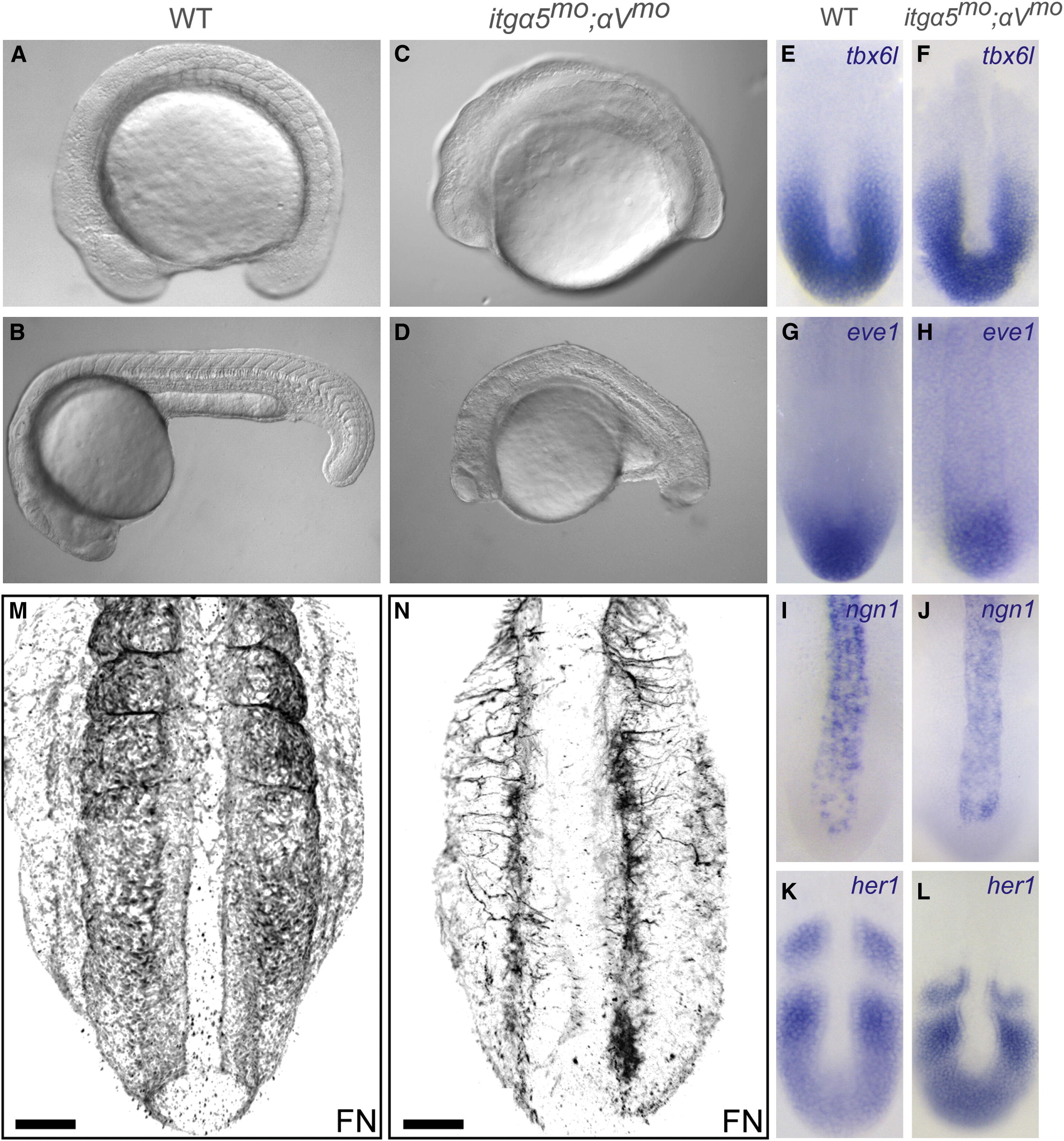
Fig. 1
Axis Elongation Defects after Loss of Both itgα5 and itgαV (A-D) Wild-type (WT) (A and B) and truncated (C and D) itgα5mo;αVmo embryos at the end of trunk elongation; i.e., 16-somite-stage embryos (A and C) and 24 hr postfertilization (hpf) (B and D). At the 16-somite stage, we find that distance from the otic vesicle to the anterior of the head in itgα5mo;αVmo embryos (n = 30) is 74% (SD = 8%; p < 0.05) of that in WT embryos (n = 20) and that the distance from the otic vesicle to the tip of the tail is 71% of that in WT embryos (SD = 7%; p < 0.05). (E–L) In situ hybridization of tail bud gene expression in 13-somite-stage embryos. (M and N) FN immunolocalization in 16-somite-stage WT (M) (n = 10) and itgα5mo;αVmo embryos (N) (n = 17). Note the reduction in FN matrix as well as the prominent medial-lateral fiber orientation in (N). Scale bars are 50 μm. In (A–D), anterior is the left. In (E–N), anterior is up. See also Figure S1./p>