
|
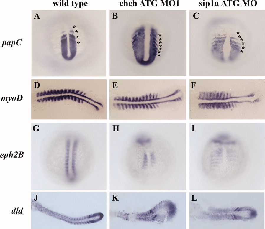
Fig. 2 Patterning of the presomitic and somitic mesoderm is disrupted in ChCh and Sip1a compromised embryos. A-L: Whole-mount (A-C,G-I) and flat-mount (D-F,J-L) RNA in situ hybridization of somite markers in wild-type, ChCh and Sip1a compromised embryos. All views are dorsal; anterior to the top (A-C,G-I) and anterior to the left (D-F,J-L). A-C: The expression domains of the PSM marker, papC is broader mediolaterally in ChCh and Sip1a compromised embryos than the wild-type siblings. The number of the papC expression stripes corresponding to the prospective and formed somites at the segmentation plate in chch and sip1a MO is higher than in wild-type siblings (compare asterisks number). D-F: The myogenic regulatory factor, myoD, is expressed in the posterior somite compartment in chch and sip1a MO injected embryos as in wild-type embryos. G-L: ephB2 and dld expression at the anterior half of the somites is reduced and diffuse. Asterisks denote (prospective) somites.