
|
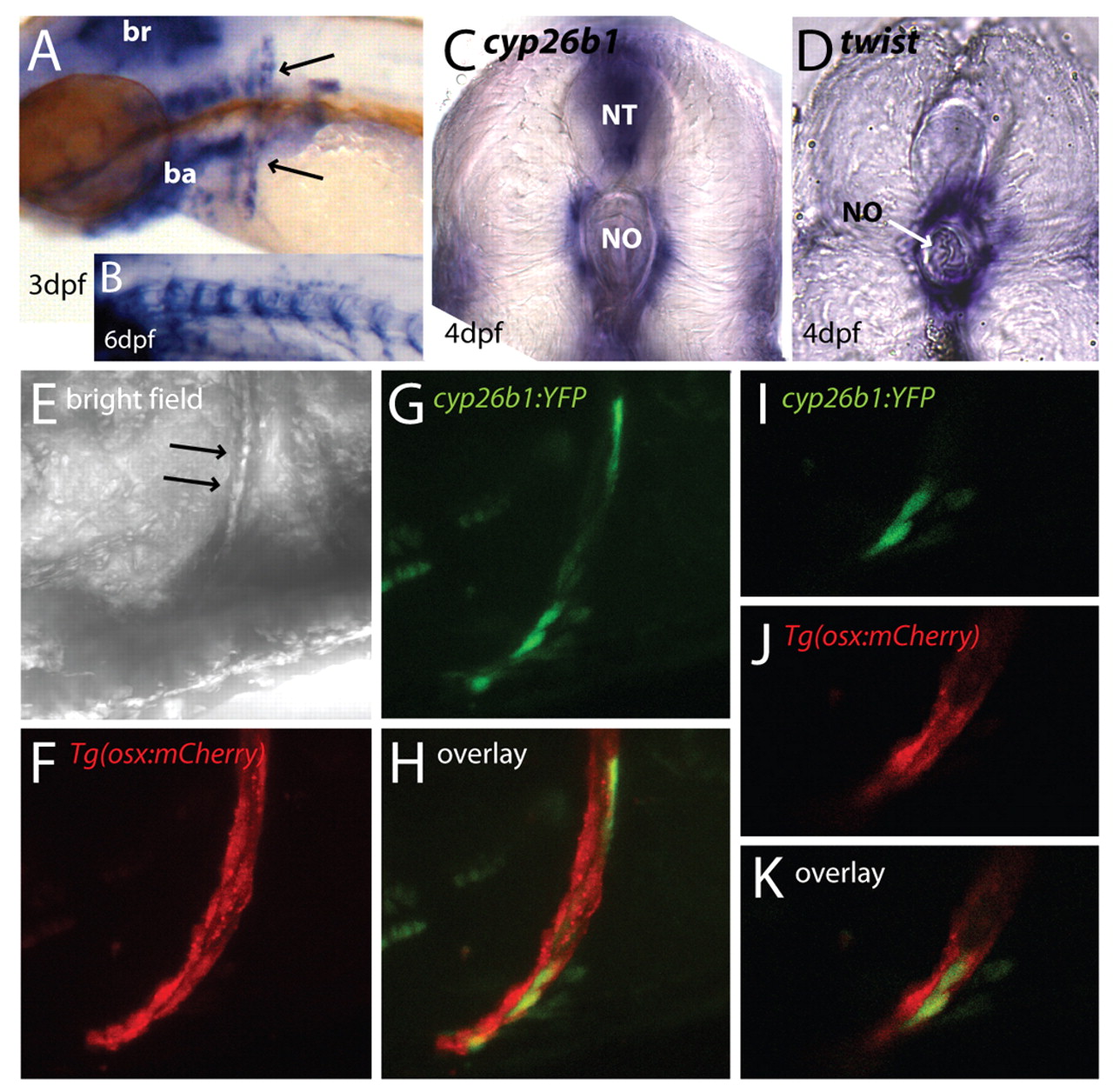
Fig. 3 cyp26b1 is expressed in zebrafish osteoblasts. (A-C) cyp26b1 expression in zebrafish. (A) Early cyp26b1 expression in zebrafish is found in the hindbrain, branchial arches, pectoral fins and the cleithrum (arrows). (B) At 6 dpf, cyp26b1 expression is seen in a segmented pattern around the notochord. A mutant embryo is shown, as cyp26b1 mRNA levels are higher in mutants than wild-type embryos. (C,D) Vibratome transverse sections (4 dpf, 100 μm) showing cyp26b1 expression (C) surrounding the notochord (no) and in the neural tube (nt), and expression of the sclerotome marker twist in cells juxtaposed to the notochord (D), similar to cyp26b1 expression. (E) Bright-field view of an embryonic trunk with arrows indicating the cleithrum. (F-K) Co-localization of cyp26b1:YFP and osx:mCherry in osteoblasts of the cleithrum. (F) Cleithrum cells are labelled in Tg(osx:mCherry) embryos. (G,H) Transient expression of cyp26b1:YFP in cells of the cleithrum at 4 dpf, with projection in H demonstrating colocalization of both genes in the same cells. (I-K) Single confocal scans of a part of the projections shown in F-H. Anterior is towards the left, except where stated otherwise.