
|
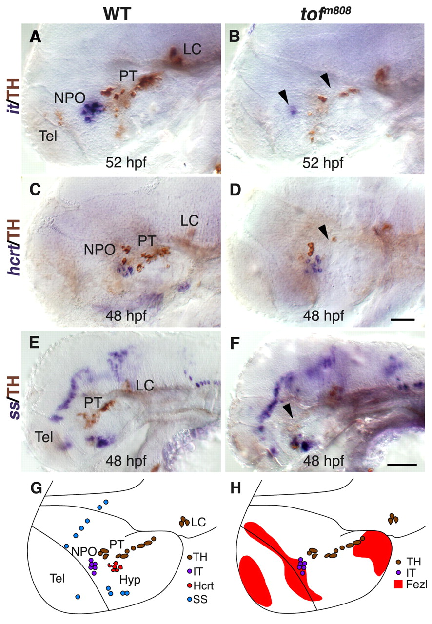
Fig. 1 Analysis of hypothalamic neurons in fezl/tofm808 mutant embryos. (A-F) Heterozygous (WT; A,C,E) embryos and their too fewm808 (tofm808) mutant siblings (B,D,F) were fixed at 52 (A,B), and 48 (C-F) hours post fertilization (hpf) and subjected to whole-mount in situ hybridization with antisense RNA probes directed against either the hypothalamic neuropeptide isotocin, which is the zebrafish ortholog of oxytocin (it; A,B), hypocretin/orexin (hcrt; C,D) or somatostatin (ss; E,F). After probe color development, all specimens were subjected to immunostaining with an anti-tyrosine hydroxylase (TH) antibody to detect DA neurons. WT heterozygous and tofm808 embryos were scored by TH staining followed by sequencing-based genotyping. Black arrowheads indicate deficiencies in DA and IT neurons in too few embryos. (G,H) SchCsup>m808 embryos were scored by TH staining followed by sequencing-based genotyping. Black arrowheads indicate deficiencies in DAematic representations of the examined hypothalamic cell types and of fezl/tof expression domains in a 2-day-old WT embryo. All panels show lateral views of the embryo, anterior to the left. Hyp, hypothalamus; LC, locus coeruleus; NPO, neurosecretory preoptic area; PT, posterior tuberculum; Tel, telencephalon. Scale bars: 50 μm in A-D; 100 μm in E,F.