
|
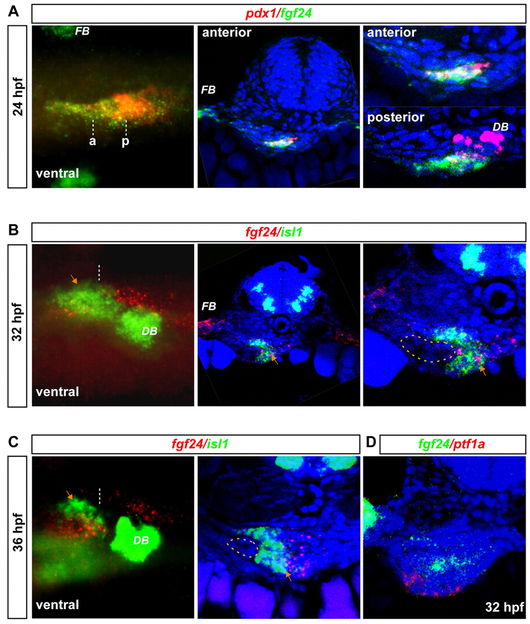
Fig. 4 fgf24 is expressed in the pancreatic endoderm and in the pancreatic LPM prior to and during ventral pancreatic bud formation. (A) fgf24 (green) expression analysis by fluorescent whole-mount in situ hybridization at 24 hpf with pdx1 probe (red). A whole-mount ventral view (epifluorescence microscopy) is shown on the left panel (anterior to the left and left side of the embryo up) with anterior (a) and posterior (p) level of section. The transverse sections were analyzed by confocal microscopy, through the anterior pancreatic domain (middle panel, global view; right panel, close-up) and more posteriorly, through the dorsal pancreatic bud (right panel, close-up). (B) Expression of fgf24 (red) compared with isl1 (green) at 32 hpf (transverse section, close-up in the right panel). Expression in the pancreatic LPM is indicated by orange arrows. (C) fgf24 and isl expression at 36 hpf. fgf24 expression appears as small red grains owing to its weak expression. (D) fgf24 (green) and ptf1a (red) expression at 32 hpf. The images of transverse sections presented in B and C are flat stacking of several consecutive optical sections. DB, dorsal pancreatic bud; FB, pectoral fin bud.