
Image
|
Figure Caption
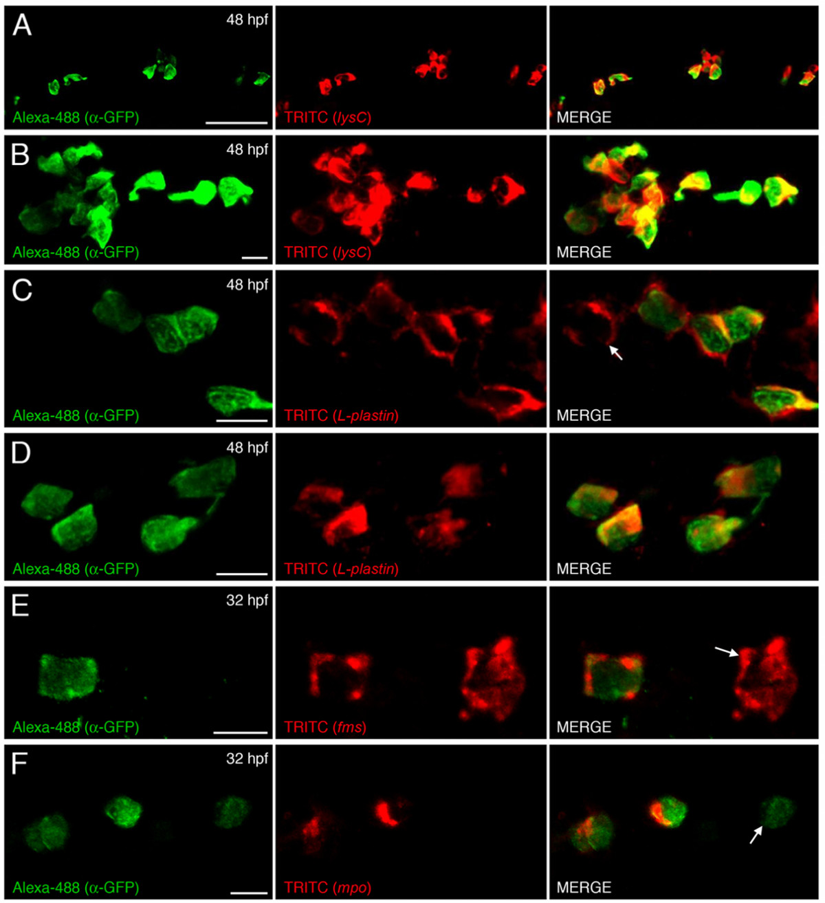
Fig. 4 EGFP-labeled cells co-express myeloid lineage markers. Dual immunohistological detection of EGFP and in situ hybridization detection of lysC (A and B), L-plastin (C and D) within 48 hpf and fms (E), mpo (F) within 32 hpf lysC::EGFP animals. (A/C/E/F and B/D) Summed Z stacks within posterior ICM compartment and within hindbrain, respectively, of transgenic animals. Arrow in C and E denotes EGFP-negative cell expressing L-plastin and fms, respectively. Arrow in F denotes EGFP-expressing cell that does not co-express mpo. Anterior to left in all images. Scale bars: 50 μm in A; 10 μm in B-F.
Figure Data
Acknowledgments
This image is the copyrighted work of the attributed author or publisher, and
ZFIN has permission only to display this image to its users.
Additional permissions should be obtained from the applicable author or publisher of the image.
Full text @ BMC Dev. Biol.