
|
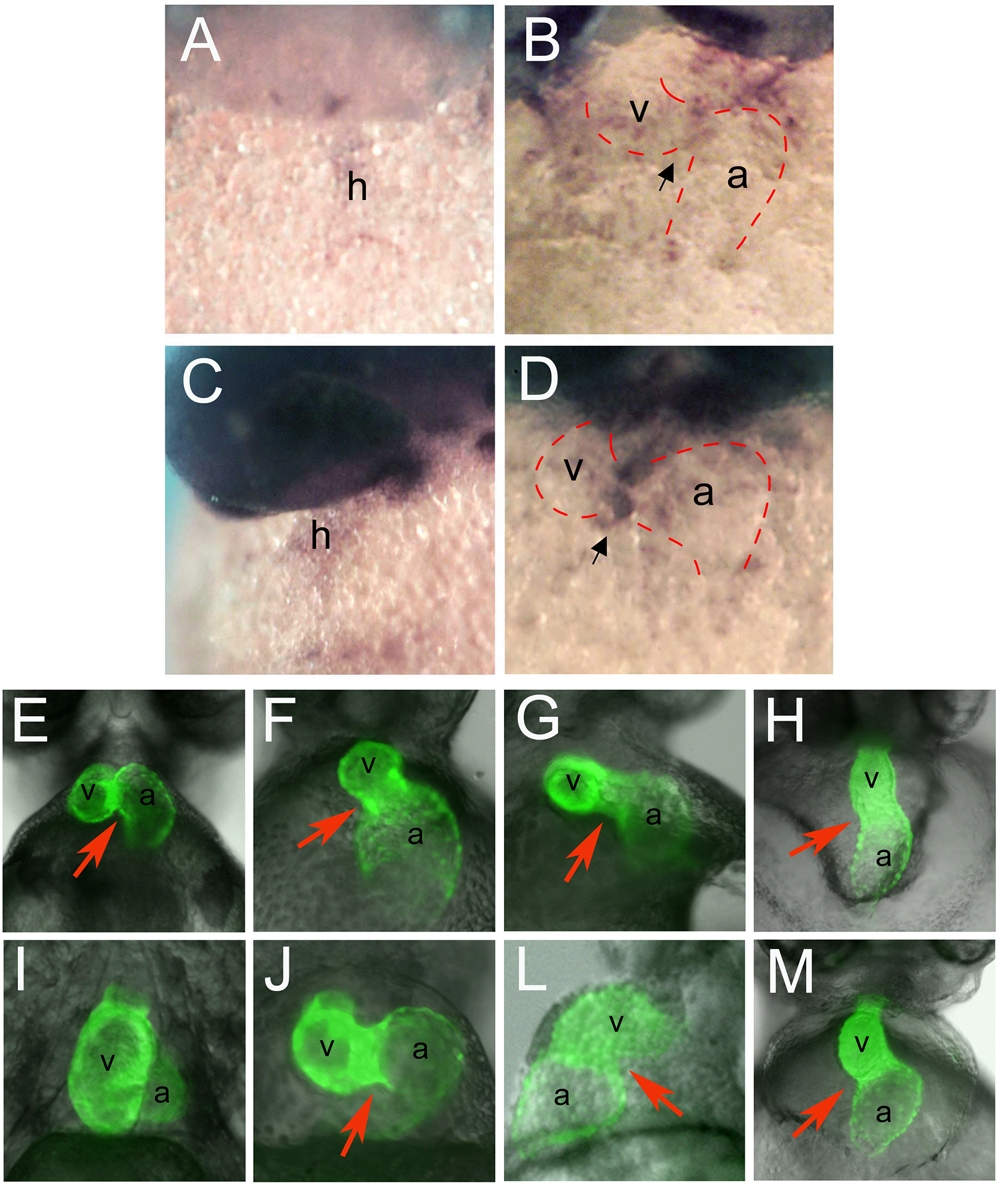
Fig. 1 (A–D) Whole mount RNA in situ hybridization of zebrafish tbx3b and tbx2a expression; ventral views show the heart in its maximum extension from the anterior to the posterior pole. Zebrafish tbx3b is expressed throughout the extent of the heart tube at 31 hpf (A) and becomes restricted to the AVC at 42 hpf (B). Zebrafish tbx2a is expressed al low levels throughout the extent of the linear heart tube at 31 hpf (C). At 42 hpf tbx2a transcripts are present in the AVC at high levels (D). Black arrow points to the AVC. (E–M) Ventral views of the heart of a mlc2a::GFP transgenic line that expresses GFP in the myocardium, at 48 hpf (E–H) and 72 hpf (I–M). (E) In wild type 48 hpf embryos, the atrium has moved upward and is positioned at the same anterior-posterior level as the ventricle; (I) later the atrium becomes localized dorsal to the ventricle at 72 hpf. (F, J) Injection of 2.5 ng of tbx3b morpholino into one-cell stage embryos results in delayed heart looping and abnormal AVC. (G, L) Injection of 10 ng of tbx2a MO results in a similar delay in heart looping and an enlargement of the AVC. (H, M) Injection of both tbx3b and tbx2a MOs, at 1.75 and 5 ng respectively, results in the failure to form the AVC constriction and absence of looping. Red arrow indicates the AVC. a, atrium; h, heart; v, ventricle.