
|
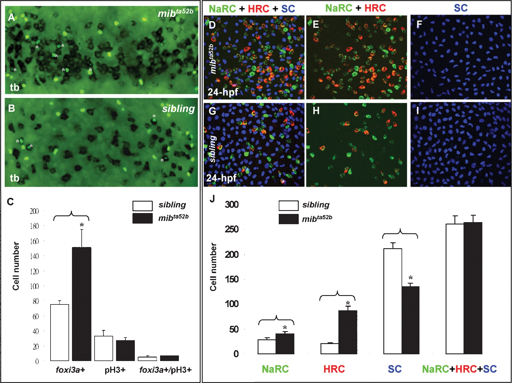
Fig. 3 Excess Epidermal Ionocytes Detected in mibta52b Occur at the Expanse of Epidermal Stem Cell Fate. (A–C) Detection of the cell proliferation activity within the epidermal ionocyte domain between mibta52b and siblings at the tail bud (tb) stage. For mibta52b embryos (B) or their siblings (A), epidermal ionocyte progenitors were labeled with foxi3a (black), and mitotic cells were labeled with phosphor-histone 3 (pH3) antibody staining (green). Cells which were double positive for foxi3a and pH3 are highlighted by asterisks. (C) Quantitative comparison of the epidermal ionocyte progenitor number, mitotic divided cell number, and mitotic divided epidermal ionocytes between mibta52b embryos and siblings. (D–I) The excess epidermal ionocytes in mibta52b embryos occur at the expense of the epidermal stem cell fate. For 24-hour post-fertilization (hpf) mibta52b embryos (D–F) and siblings (G–I), differentiating Na+,K+-ATPase-rich cells (NaRCs), H+-ATPase-rich cells (HRCs), and epidermal stem cells were labeled with atp1b1b (green), ca2a, (red), and P63 (blue) staining, respectively. (J) Quantitative comparison of NaRC, HRC, and epidermal stem cell numbers between mibta52b embryos and siblings. The cell number in (C and J) is presented as the mean±S.D. *, p<0.05, compared with siblings, as determined by Student's t-test.