
|
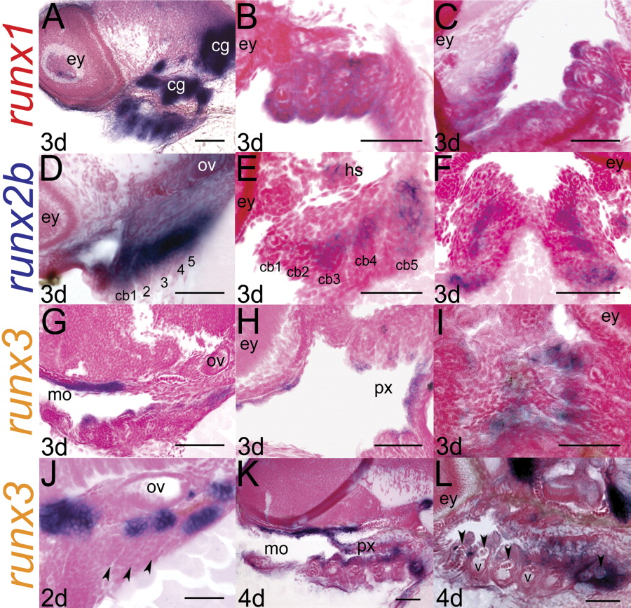
Fig. 3 Analysis of tissue sections confirms compartmentalized localization of runx transcripts in the pharyngeal domain. (A-C) runx1; (D-F) runx2b; (G-L) runx3; (A,B,G,J-L) sagittal sections, anterior to left; (B,E,H) frontal sections, anterior to left; (C,F,I) frontal sections, anterior to top. A sagittal section through a 3dpf embryo reveals runx1 expression in pharyngeal arches (A). Frontal sections verify staining in the entire epithelial layer of the pharyngeal arches (B,C). runx2b expression is restricted to mesenchymal cells deep in the core of the arches (D-F), while runx3 marks the endodermal epithelium (G-I) in 3dpf larvae. This endodermal expression of runx3 is present at 2dpf in the pouch endoderm (J), and later in the endodermal lining of the mouth and pharynx (K, L). At 4dpf, runx3 is expressed in differentiating chondrocytes (arrowheads, L). cb, ceratobranchial (numbers denote cb identity); cg, cranial nerve ganglia; hs, hyosymplectic; mo, mouth; px, pharynx; ov, otic vesicle; v, blood vessel. Scale bars = 50 μm.