
|
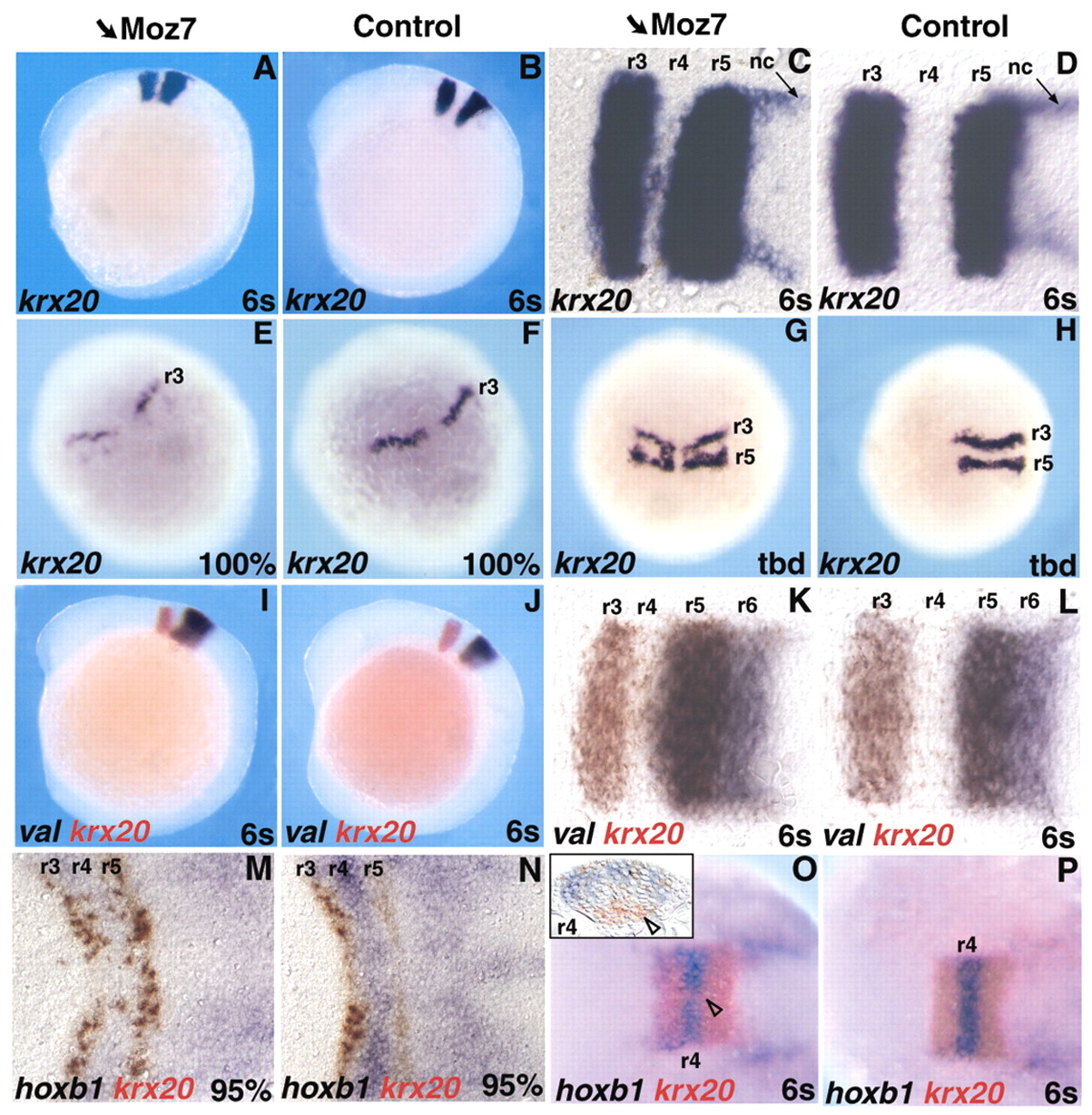
Fig. 3 Knocking-down iro7 results in an anterior expansion of r5 at the expense of r4. (A-P) Whole-mount in situ hybridisation with the probes indicated (bottom left of each picture, colour coded) on embryos injected with Moz7 (A,C,E,G,I,K,M,O) or control embryos (B,D,F,H,J,L,N,P). Anterior is towards the left, except in E-H where anterior is towards the top. The inset in O presents a transverse section at the level of r4. The arrowheads indicate a group of cells expressing krx20 ectopically in the ventral part of r4. (C,D,K,L,M,N) Dorsal views of flat-mounted embryos. Stages are indicated at the bottom right of each picture. nc, neural crest.