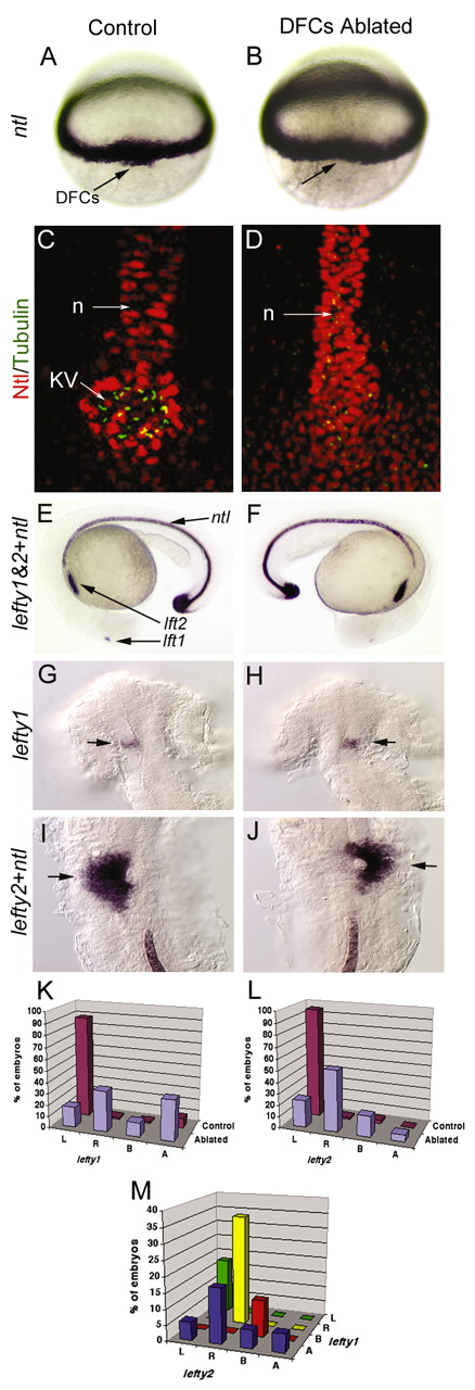
Fig. 2 Laser ablation of DFCs alters LR patterning without affecting development of the midline. Unablated control embryos expressed ntl in DFCs (arrow) at 60% epiboly (A) and in the notochord at 24 hours post-fertilization (E). Laser ablation eliminated DFCs (arrow, dorsal view at 60% epiboly) but did not alter ntl expression in equatorial mesoderm (B) or in notochord and tailbud at 24 hpf (F). Control embryos (C) and DFC-ablated embryos (D) were immunostained with anti-acetylated Tubulin antibodies (green) to detect cilia and anti-ntl antibodies (red) to detect the notochord (n) and Kupffers vesicle (KV). Both control and DFC-ablated embryos showed contiguous ntl staining in the notochord, even in embryos (n=2/6) that failed to form KV (D). (G-J) In control embryos, we observed normal expression of lft1 (arrow) in the left dorsal diencephelon (G) and lft2 (arrow) in the left heart primordia (I). In DFC-ablated embryos, lft1 (arrow) in the diencephalon (H) and lft2 (arrow) in the heart primordia (J) were frequently reversed. (K-M) Analysis of lft1 expression in the diencephalon (K) and lft2 expression in the heart primordia (L) in control (n=71) and DFC-ablated (n=17) embryos. (M) Analysis of lft1 expression plotted against lft2 expression in DFC-ablated embryos indicated that the predominant class of DFC-ablated embryos displayed reversal of both brain and heart markers (yellow bar). L, left; R, right; B, bilateral; A, absent gene expression.
Image
Figure Caption
Figure Data
Acknowledgments
This image is the copyrighted work of the attributed author or publisher, and
ZFIN has permission only to display this image to its users.
Additional permissions should be obtained from the applicable author or publisher of the image.
Full text @ Development

