
|
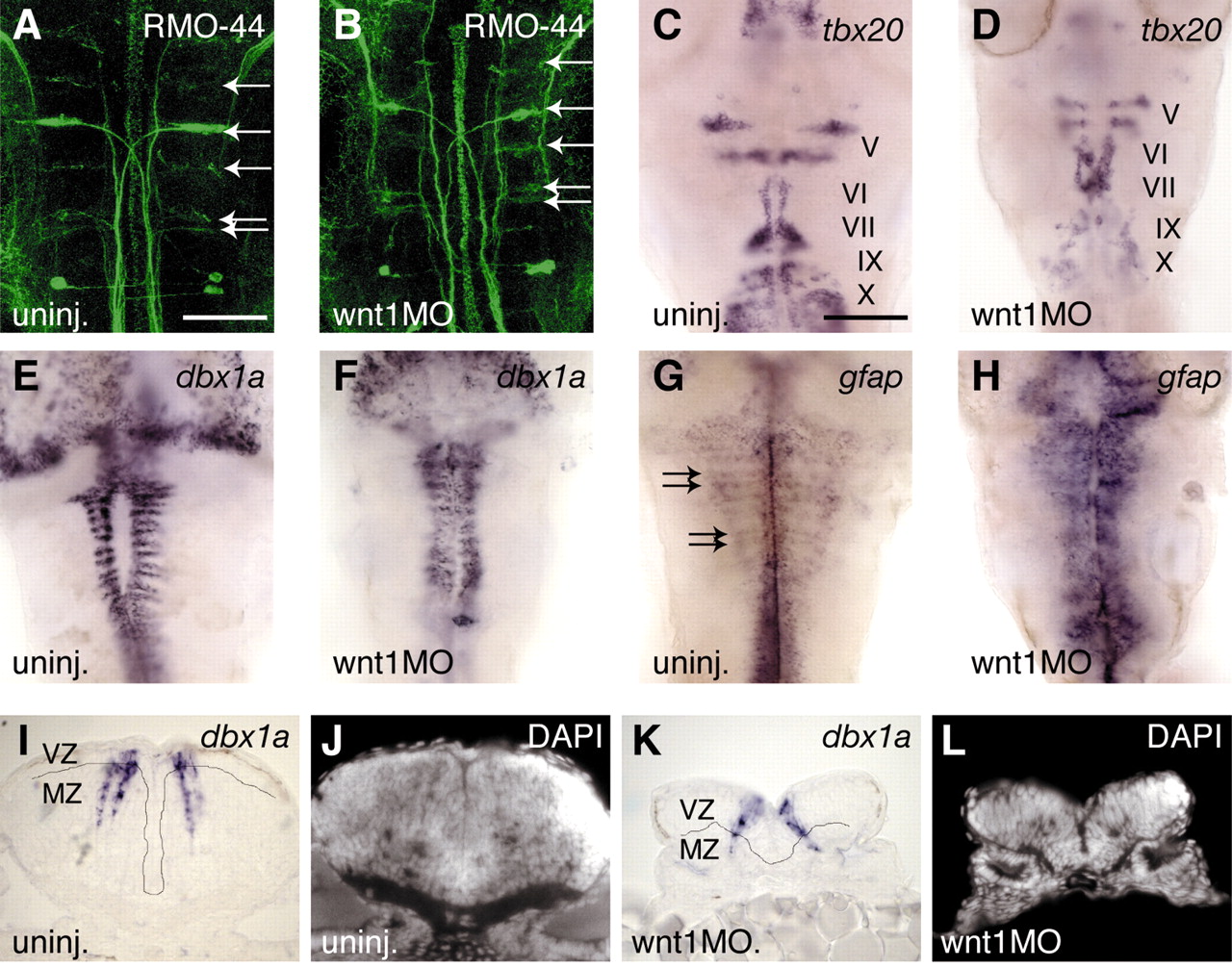
Fig. 5 Neurogenesis at 48 hours in wnt MO-injected embryos. (A,B) Reticulospinal neurons labelled by RMO-44 antibody (arrows) are still present (11/14 embryos), and cranial motor nerves (V, VI, VII, IX and X) labelled with tbx20 (C,D) are mildly hypomorphic (11/11 embryos). The number of dbx1a-expressing cells appears to be similar in uninjected and morphant embryos (E,F), but their distribution in stripes adjacent to boundaries is disrupted (20/20 embryos). Sections of uninjected (I,J) and wnt1MO embryos (K,L) show that in uninjected embryos there are many neurons expressing dbx1a in the mantle zone (MZ); in the morphants, most dbx1a expression occurs in the ventricular zone (VZ) of progenitors (4/4 embryos), as determined by DAPI stain (J,L) and by absence of Hu staining (not shown). (G,H) gfap expression occurs in stripes adjacent to boundaries in uninjected embryos (arrows in G), and ectopically in wnt1 MO embryos (H, 12/13 embryos), suggesting that neural progenitors have switched fate to glial cells. Scale bar: in A, 50 �m for A,B,I-L; in C, 100 �m for C-H.