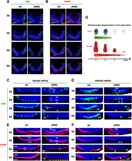
Rods undergo apoptotic-like degeneration in pday mutants, whereas cones progressively shrink. (A,B) Images of wild-type and pday mutant retinas carrying the transgenes Tg[rho:NLS-eGFP] and Tg[gnat2:NLS-tdTomato], which visualize rods (green, A) and cones (red, B), respectively. Nuclei were counterstained with Hoechst 33342 (blue). Rectangles indicate the dorsal and ventral ONLs, shown in C-F. (C) The dorsal ONL of wild-type and pday mutant retinas with Tg[rho:NLS-eGFP] (green). In pday mutants, rods had condensed, round nuclei (white arrowheads) at 3 dpf, and rods were drastically reduced in number at 4 dpf and disappeared at 5 and 6 dpf (asterisks), except in the peripheral region near the dorsal ciliary marginal zone (CMZ). pe, retinal pigment epithelium. (D) The ventral ONL of wild-type and pday mutant retinas with Tg[rho:NLS-eGFP] (green). In pday mutants, rods had condensed, round nuclei (white arrowheads) at 3 and 4 dpf white dotted lines. In pday mutants, rods disappeared at 5 and 6 dpf (asterisks), except in the peripheral region near the ventral CMZ. (E) The dorsal ONL of wild-type and pday mutant retinas with Tg[gnat2:NLS-tdTomato] (red). In wild type, cones formed a monolayer at 3 dpf and two subnuclear layers after 4 dpf (white dotted lines). In pday mutants, tdTomato-negative nuclei were observed, the positions of which were identical to those of condensed rod nuclei (white arrowheads). From 4 to 5 dpf, cones shrunk progressively and became flattened (yellow line). At 6 dpf, cones were extremely flattened and disappeared, leaving gaps (yellow arrowheads), except in the peripheral region near the dorsal CMZ. (F) The ventral ONL of wild-type and pday mutant retinas with Tg[gnat2:NLS-tdTomato] (red). In wild type, cones differentiated at 3 dpf, but progressively disappeared toward the periphery of the ventral ONL, where rods densely differentiated. In pday mutants, tdTomato-negative round nuclei appeared at the place where pyknotic-like rod nuclei were observed (white arrowheads, D) at 3 dpf, and ONL thickness was progressively decreased after 4 dpf. Interestingly, after 5 dpf, the cone differentiating area was expanded toward the periphery of the ventral ONL (asterisks), where rods disappeared. (G) Cone and rod degeneration process in pday mutants. Rods undergo acute degeneration with apoptotic-like pyknotic nuclei during 3-4 dpf, whereas cones undergo progressive shrinkage of cell volume during 4-6 dpf and disappear by 6 dpf. Sample sizes are shown in Table S3. Scale bars: 40 µm (A,B).
|
