|
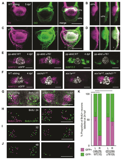
Loss of function of cachd1 disrupts habenular efferent connectivity, is epistatic to removal of the parapineal signal, and causes precocious neurogenesis. (A and C) Dorsal views and (B and D) sagittal projections (dorsal left) of the IPN showing DiI (magenta) and DiD (green) labelling of left- and right-sided habenula neuron axon terminals predominantly innervating the dIPN and vIPN respectively, and raphe (r), in 5 dpf wild-type [(A) and (B), n = 3] or cachd1u761 mutant [(C) and (D), n = 8] larvae. (E) Dorsal views of 4 dpf wild-type or cachd1u761 mutant epithalami in which the parapineal was ablated before leftward migration (pineal complex marked by zf104Tg, u711Tg alleles with antibody to GFP; white) after double FISH with kctd12.1 (magenta; n = 26 of 29 wild-type siblings, 11 of 12 cachd1u761 mutants) or kctd12.2 (green; n = 19 of 23 wild-type siblings, 5 of 5 cachd1u761 mutants). (F) Dorsal views of 4 dpf larvae from a cross of carriers of cachd1u761 and sox1aups8 alleles after FISH with kctd12.1 [magenta; pineal complex as (C), white]. n = 4 wild-types, 3 cachd1u761 mutants, 4 sox1aups8 mutants, 3 sox1aups8, cachd1u761 double mutants. (G) Dorsal views of Et(gata2a:EGFP)pku588 wild-type or cachd1u761 mutant habenulae incubated at 48 hours post fertilization (hpf) with a pulse of BrdU to label newly born neurons, then processed for immunohistochemistry at 5 dpf with antibody to GFP (magenta) and antibody to BrdU (green). DAPI (4′,6-diamidino-2-phenylindole) counterstain marks nuclei (gray). (H to J) Segmentation of confocal stacks from Et(gata2a:EGFP)pku588 wild-type or cachd1u761 mutant larvae incubated at (H) 24, (I) 32 and (J) 48 hpf with a pulse of BrdU, then processed at 5 dpf as in (G). Double-positive cells are represented in magenta; BrdU-positive only cells are indicated in green. Times of pulse indicated at top right. (K) Quantification of the proportion of BrdU-positive neurons that also expressed Et(gata2a:EGFP)pku588 (magenta) in 5 dpf wild-type or cachd1u761 larvae incubated with a pulse of BrdU at 24 hpf (all timepoints presented in fig. S11). Error bars represent 95% confidence intervals. Total number of cells and larvae for each genotype indicated in axis label in parentheses. Q′ test of equality of proportions [24 hpf, degrees of freedom (DF) = 3, χ2 = 40.94, P = 6.7 × 10⁻⁹], post hoc pairwise comparisons using a modified Marascuilo procedure with Benjamini-Hochberg correction for multiple testing, **** P ≤ 0.005. Scale bars, 50 µm in (A) to (H).
|