|
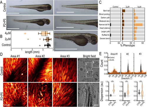
Peptides induce phenotypic changes in zebrafish consistent with TANGO1/cTAGE5 inhibition. A Zebrafish larvae at 3 dpf. Fish were treated with P2 + P5 (4 μM), or left untreated in E3 (control). Magnified images of the tail and yolk on the right. Scale bars 1 mm. B Quantification of larva length from head to tail (boxplot with median and interquartile range N = 21, 19, 23 in control, 2 μM and 4 μM respectively). Whiskers connect the maximum and minimum. C Quantification of phenotypes observed in fish for n = 21, 19, 23 replicates in control, 2 μM and 4 μM respectively, from three independent experiments. D Maximum projection of the entire Z-stack of SHG images from three different tail fin regions (Areas #1, 2, 3) as indicated in the bright-field images. Images were acquired from three control and treated fish each, with approximately similar regions of the tail chosen as Area #1, 2, and 3. Scale bars 100 μm. E Quantification of collagen fiber direction obtained from the SHG signal in the three segmented areas approximately represented in (D) Top row: Collagen fiber direction histograms from a representative control or treated fish. Bottom row: Collagen fiber dispersion distribution plots from a representative control or treated fish. This measure represents the dispersion of the preferred orientation of collagen fibers in the image. Control measurements are represented in black and P2 + 5 (4 μM) is treated in orange. Kolmogorov–Smirnov test *** p < 0.001.
|