IMAGE

Fig. 2

ID
ZDB-IMAGE-240426-86
Source
Figures for Raote et al., 2024
Image
Figure Caption

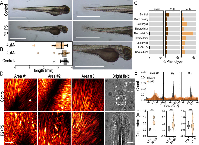
Fig. 2 Peptides induce phenotypic changes in zebrafish consistent with TANGO1/cTAGE5 inhibition.

A Zebrafish larvae at 3 dpf. Fish were treated with P2 + P5 (4 μM), or left untreated in E3 (control). Magnified images of the tail and yolk on the right. Scale bars 1 mm. B Quantification of larva length from head to tail (boxplot with median and interquartile range N = 21, 19, 23 in control, 2 μM and 4 μM respectively). Whiskers connect the maximum and minimum. C Quantification of phenotypes observed in fish for n = 21, 19, 23 replicates in control, 2 μM and 4 μM respectively, from three independent experiments. D Maximum projection of the entire Z-stack of SHG images from three different tail fin regions (Areas #1, 2, 3) as indicated in the bright-field images. Images were acquired from three control and treated fish each, with approximately similar regions of the tail chosen as Area #1, 2, and 3. Scale bars 100 μm. E Quantification of collagen fiber direction obtained from the SHG signal in the three segmented areas approximately represented in (D) Top row: Collagen fiber direction histograms from a representative control or treated fish. Bottom row: Collagen fiber dispersion distribution plots from a representative control or treated fish. This measure represents the dispersion of the preferred orientation of collagen fibers in the image. Control measurements are represented in black and P2 + 5 (4 μM) is treated in orange. Kolmogorov–Smirnov test *** p < 0.001.

Acknowledgments
This image is the copyrighted work of the attributed author or publisher, and ZFIN has permission only to display this image to its users. Additional permissions should be obtained from the applicable author or publisher of the image. Full text @ Nat. Commun.