
Fig. 4
- ID
- ZDB-FIG-240405-5
- Publication
- Lin et al., 2023 - Biallelic COQ4 Variants in Hereditary Spastic Paraplegia: Clinical and Molecular Characterization
- Other Figures
- All Figure Page
- Back to All Figure Page
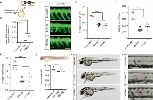
Functional characterization of coq4 in hb9::GFP zebrafish. (A) Schematic for generating coq4-KD larvae. Two coq4-specific MOs (morpholino oligomer, red line) targeting the first translation initiation site (ATG-MO) or the 3′ splicing site in exon 4 (E4I4-MO) were microinjected into fertilized, one-cell-stage zebrafish embryos. (B) QPCR (Quantitative PCR) analysis of coq4 mRNA levels in zebrafish embryos microinjected with control-MO or coq4-MO at 3 dpf (days post fertilization) (n = 4, one-way ANOVA [analysis of variance]). (C) Motor axon growth of hb9::GFP zebrafish embryos at 2 dpf. Scale bar, 100 μm. (D) Average length of motor axons in (C) (n = 10 zebrafish/group, about 30 axons per fish were examined; one-way ANOVA). Locomotor performance was assessed by (E) distance covered and (F) average velocity in coq4-MO larvae at 5 dpf (n = 10 zebrafish/group, one-way ANOVA). (G) Smaller head region (enclosed by dashed line) of coq4-MO larvae compared to control-MO larvae at 6 dpf (n = 10 zebrafish/group, one-way ANOVA). (H, I) Pericardial edema formation (black arrow; H) and U-shaped somites (red arrow; I) in coq4-KD larvae but not in control-MO larvae (blue arrow, chevron-shaped somites) at 2 dpf. [Color figure can be viewed at wileyonlinelibrary.com] |