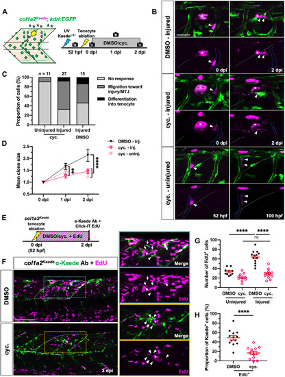
Fig. 9
|
. Model of fibroblast plasticity and tendon regeneration in zebrafish. (A) Schematic showing distinct plasticity of different sclerotome-derived fibroblast populations in the zebrafish trunk at 52 hpf, including progenitor-like perivascular/interstitial fibroblasts, as well as specialized tenocytes, fin mesenchymal cells, and notochord-associated fibroblasts. During normal development, perivascular fibroblasts act as pericyte precursors, while interstitial fibroblasts function as tenocyte progenitors. By contrast, both perivascular and interstitial fibroblasts contribute to tenocyte regeneration upon tendon injury, while perivascular fibroblast–derived pericytes as well as specialized fibroblasts do not exhibit such regenerative plasticity. (B) Timeline of fibroblast response to tenocyte ablation. After tenocyte ablation, perivascular fibroblast–mediated tenocyte regeneration proceeds in three partially overlapping phases. First, perivascular fibroblasts are recruited to the injured MTJ from the vasculature between 0 and 1 dpi. Second, the activated fibroblasts then undergo one to two rounds of cell division in a Hh signaling–dependent manner starting at 1 dpi and concurrently up-regulate tenocyte marker genes, prelp and tnmd. Last, by 4 dpi, a subset of these differentiating fibroblasts matures into new tenocytes extending characteristic cellular processes into the newly regenerated MTJ. |

