
Fig. 4
- ID
- ZDB-FIG-230719-31
- Publication
- Salehin et al., 2022 - Ventricular Anisotropic Deformation and Contractile Function of the Developing Heart of Zebrafish in-vivo
- Other Figures
- All Figure Page
- Back to All Figure Page
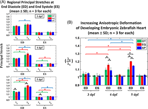
(A) Regional principal stretches λ1, λ2 at end-diastole (ED) and end-systole (ES) during a cardiac cycle at the outflow (OT), equatorial (EQ), and apex (AP) regions of developing zebrafish heart at 3-, 4-, and 5-dpf where λ2 denote principal stretches near the latitudinal direction and λ1 denote that at the direction perpendicular from the former. From 4-, 5-dpf groups, at ED, we noted greater deformation (designated as λ2) at the direction near the ALR in each of OT, EQ, and AP regions. Same were found at ES state indicating their anisotropic deformation, except in AP from 4-dpf. From 3-dpf group, we noted only slightly anisotropic with significantly higher λ2 at AP region. Statistically significant differences (P < 0.05) are denoted by asterisk. (B) Ratios of principal stretch components (λ2/λ1) at ED and ES states from outflow (OT), equatorial (EQ), and apex (AP) regions. Statistically significant differences (P < 0.05) are denoted by asterisk |