
Fig. 5
- ID
- ZDB-FIG-230519-39
- Publication
- He et al., 2022 - Translational control by maternal Nanog promotes oogenesis and early embryonic development
- Other Figures
- All Figure Page
- Back to All Figure Page
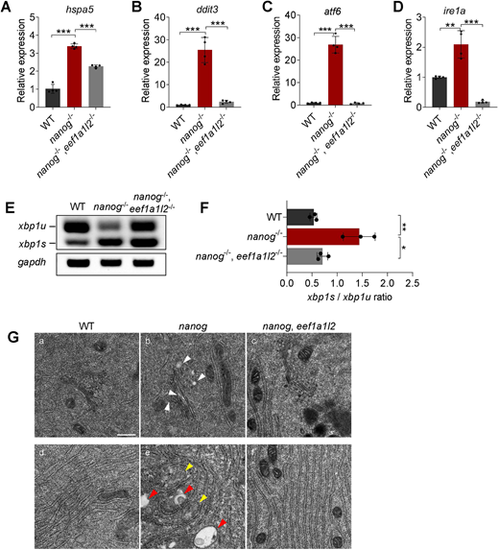
Depletion of eef1a1l2 ameliorates ER stress and UPR in nanog mutant oocytes. (A-D) RT-qPCR analysis showing decreased expression of hspa5 (A), ddit3 (B), atf6 (C) and ire1a (D) in nanog and eef1a1l2 double-mutant ovaries, compared with nanog mutant ovaries. **P<0.01, ***P<0.001. n=4. (E,F) RT-PCR examination of xbp1 splicing. The ratio of spliced xbp1 (xbp1s) mRNA to unspliced xbp1 (xbp1u) mRNA was increased in nanog mutant ovaries, but restored in nanog and eef1a1l2 double-mutant ovaries. gapdh was used as internal control. The xbp1s/xbp1u ratio in F represents the intensity ratio of the corresponding PCR product bands in E. *P<0.05, **P<0.01. n=3. (G) ER, Golgi and mitochondria structure in WT, nanog mutant and double-mutant stage I oocytes as revealed by transmission electron microscopy. White arrowheads indicate Golgi apparatus, yellow arrowheads indicate mitochondria, red arrowheads indicate lysosomes. Scale bar: 0.5 μm. |