
Fig. 7
- ID
- ZDB-FIG-230126-23
- Publication
- do Amaral et al., 2022 - The perinuclear region concentrates disordered proteins with predicted phase separation distributed in a 3D network of cytoskeletal filaments and organelles
- Other Figures
- All Figure Page
- Back to All Figure Page
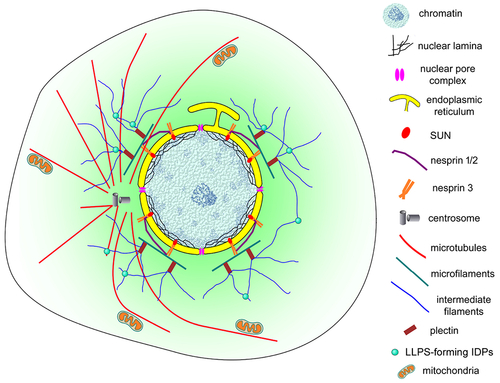
Schematic representation of the nuclear cloud of eukaryotic cells. This model illustrates the complex organization of the perinuclear space (shown in light green) of eukaryotic cells. An intricate 3D network of cytoskeletal filaments is found attached to the nuclear membrane (yellow). Several proteins participate in this organization: cytoskeletal (microfilaments in green, microtubules in red and intermediate filaments in blue) and cytoskeletal associated proteins (plectin, alpha-actinin, and others), nesprins, SUNs, among many others. Dynamic biomolecular condensates drove by intrinsically disordered proteins (IDPs), have a higher propensity to be assembled (small green spheres). Different organelles and cellular structures are also present in the nuclear could: mitochondria (orange), lysosomes, endoplasmic reticulum, Golgi, vesicles, centrosome (grey), and myofibers (in muscle cells). |
Reprinted from Biochimica et biophysica acta. Molecular cell research, 1869, do Amaral, M.J., de Andrade Rosa, I., Andrade, S.A., Fang, X., Andrade, L.R., Costa, M.L., Mermelstein, C., The perinuclear region concentrates disordered proteins with predicted phase separation distributed in a 3D network of cytoskeletal filaments and organelles, 119161, Copyright (2022) with permission from Elsevier. Full text @ BBA Molecular Cell Research