Fig. 7
- ID
- ZDB-FIG-230112-41
- Publication
- Klingbeil et al., 2021 - Corpuscles of Stannius development requires FGF signaling
- Other Figures
- All Figure Page
- Back to All Figure Page
|
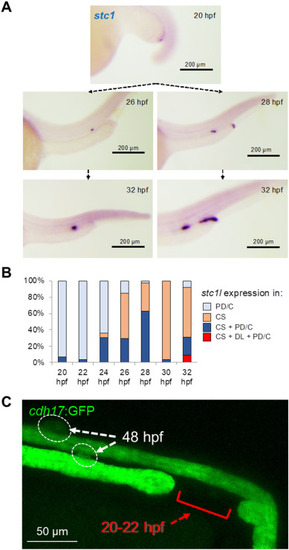
Expression of stc1 in the distal pronephros does not contribute to CS formation. (A) Analysis of stc1 expression at different developmental stages revealed natural variability in the stc1 expression. At 20 hpf stc1 was expressed in the distal pronephric duct (PD) and cloaca region. In some embryos stc1 expression was subsequently confined to the CS at later stages (left panel). However, in other embryos stc1 was also expressed outside of the CS, including the distal late tubule (DL). (B) The graph shows the percentage of embryos with stc1 expression in pronephric duct/cloaca (PD/C), corpuscles of Stannius (CS), corpuscles of Stannius and pronephric duct/cloaca (CS + PD/C), corpuscles of Stannius and distal late tubule, and pronephric duct/cloaca (CS + DL + PD/C). At 32 hpf, more than 30% of embryos expressed stc1 outside of the CS. (C) To exclude the possibility that pronephric duct cells expressing stc1 are recruited to the CS, the pronephric duct was disrupted by a 30 μm laser-induced injury at 20–22 hpf. Normal CS development occurred in all injured embryos that did not repair the gap (n = 7). |
Reprinted from Developmental Biology, 481, Klingbeil, K., Nguyen, T., Fahrner, A., Guthmann, C., Wang, H., Schoels, M., Lilienkamp, M., Franz, H., Eckert, P., Walz, G., Yakulov, T.A., Corpuscles of Stannius development requires FGF signaling, 160-171, Copyright (2021) with permission from Elsevier. Full text @ Dev. Biol.

