
Fig. 3
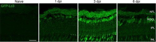
Fig. 3. Autophagy visualization at early time points after optic nerve injury using retinal cryosections of Tg(CMV:GFP-Lc3) autophagy reporter fish. The intensity of the GFP-Lc3 signal increased in the NFL and RGCL early after ONC, and peaked at 3 dpi. In addition, autophagy was observed in the IPL, in the form of GFP-Lc3+ fibers, and as a general overall increase of GFP fluorescence throughout this layer, both most pronounced at 3 dpi. In addition, GFP-Lc3+ somas inside the INL were also visible at this time point. Scale bar = 25 µm. Representative images of n = 4. Dpi, days post-injury; GFP, green fluorescent protein; INL, inner nuclear layer; IPL, inner plexiform layer; Lc3, microtubule-associated protein 1A/1B-light chain 3; NFL, nerve fiber layer; RGCL, retinal ganglion cell layer. |