
Fig. 5
- ID
- ZDB-FIG-220923-82
- Publication
- Williams et al., 2022 - Assessment of endocytic traffic and Ocrl function in the developing zebrafish neuroepithelium
- Other Figures
- All Figure Page
- Back to All Figure Page
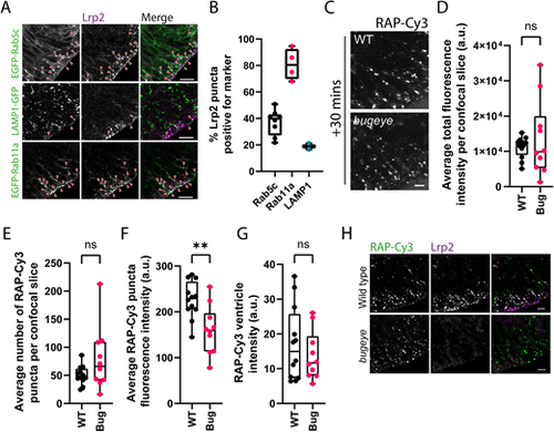
Analysis of neuroepithelial endocytic uptake of RAP in lrp2 knockout embryos. (A) Tissue sections collected from embryos transiently expressing EGFP-tagged Rab5c or Rab11a, or LAMP1–GFP at 28 hpf and stained with antibodies against Lrp2 to visualise Lrp2 localisation within the zebrafish neuroepithelium. Arrowheads point to examples of co-localisation between Lrp2 and the indicated marker. Scale bars: 10 µm. (B) Quantification of Lrp2 co-localisation with EGFP-tagged Rab5c (n=8) or Rab11a (n=4) or LAMP1–GFP (n=2). Each datapoint represents quantification from one individual embryo. Error bars show the s.d. (C) Assessment of endocytic uptake of RAP in lrp2 knockout bugeye embryos. Representative confocal microscopy images of live WT or lrp2-null zebrafish embryos following hindbrain ventricle injection of RAP–Cy3 imaged 30 min post injection. Scale bar: 5 µm. (D–G) Quantification of the average total fluorescence intensity per confocal slice (D), the average number of RAP–Cy3 puncta per confocal slice (E), the average RAP-Cy3 punctum fluorescence intensity (F) and RAP–Cy3 ventricle intensity (G) at 30 min post hindbrain injection of RAP–Cy3 in WT (n=12) and lrp2 knockout (n=10) embryos. (H) Tissue sections through the zebrafish hindbrain from WT or lrp2 knockout bugeye embryos injected with RAP–Cy3, fixed 30 min post injection and stained with antibodies against Lrp2. Scale bars: 10 µm. a.u. arbitrary units. ns, not significant; **P<0.01. Statistical comparisons between groups were made using two-tailed unpaired Student's t-test. |
| Fish: | |
|---|---|
| Observed In: | |
| Stage: | Prim-5 |