
FIGURE
Fig. 4
- ID
- ZDB-FIG-220905-20
- Publication
- Waldmann et al., 2021 - The role of Gdf5 in the development of the zebrafish fin endoskeleton
- Other Figures
- All Figure Page
- Back to All Figure Page
Fig. 4
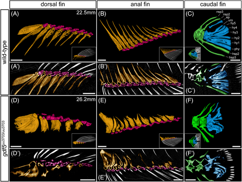
gdf5uu3703/uu3703 display defects in median fin skeletal elements. 90 dpf nonsibling wild-type (A-C), and gdf5uu3703/uu3703 (D-F) lateral views of the dorsal, anal, and caudal fin skeleton following μCT scanning and 3D segmentation (anterior to left). Fin rays (white) were removed for clarity and shown instead in the insets. (A′-F′) False-colored virtual thin sections of median fin skeletal elements. (A′,B′,D′,E′) 20 μm sections, (C′,F′) 30 μm sections. Proximal radials are colored in orange, distal radials in pink, epural, hypurals, and parhypural in blue, and preurals in green. ep, epural; hsp, hemal spine; hy, hypural; nsp, neural spine; phy, parhypural; uro, urostyle. Scale bars: 500 μm
|
Expression Data
Expression Detail
Antibody Labeling
Phenotype Data
| Fish: | |
|---|---|
| Observed In: | |
| Stage: | Adult |
Phenotype Detail
Acknowledgments
This image is the copyrighted work of the attributed author or publisher, and
ZFIN has permission only to display this image to its users.
Additional permissions should be obtained from the applicable author or publisher of the image.
Full text @ Dev. Dyn.