
FIGURE
Fig. 3
- ID
- ZDB-FIG-220905-19
- Publication
- Waldmann et al., 2021 - The role of Gdf5 in the development of the zebrafish fin endoskeleton
- Other Figures
- All Figure Page
- Back to All Figure Page
Fig. 3
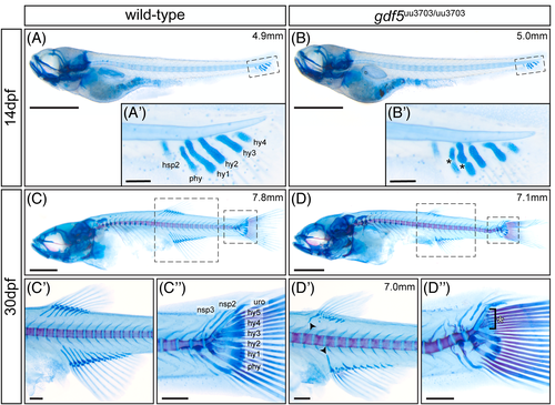
Skeletal staining reveals abnormalities in median fin skeletal organization in gdf5uu3703/uu3703 zebrafish. Lateral views of cartilage- and bone-stained wild-type fish at 14 dpf (A,A′) and 30 dpf (C,C′,C″); and gdf5uu3703/uu3703 fish at 14 dpf (B,B′) and 30 dpf (D,D′,D″). Dashed boxes (A-D) mark magnified regions (A′-D″). (B′) gdf5uu3703/uu3703 larvae display separation within the parhypural and hypural 1 cartilage condensations (black asterisks). (D′) Dorsal and anal fin radials are truncated in gdf5uu3703/uu3703 fish. The most anterior proximal radials tend to be less affected in both dorsal and anal fin (black arrowheads). This image is from a different mutant fish than displayed in (D). (D″) gdf5uu3703/uu3703 fish display truncated and slightly deformed and truncated hemal spines, parhypural and hypural 1. Hypural 3-5 are severely shortened in size (white asterisk). hsp, hemal spine; hy, hypural; nsp, neural spines; phy, parhypural; uro, uroneural. Scale bars: 1 mm (A-D), 100 μm (A′,B′) and 250 μm (C′-D″)
|
Expression Data
Expression Detail
Antibody Labeling
Phenotype Data
| Fish: | |
|---|---|
| Observed In: | |
| Stage Range: | Days 14-20 to Days 30-44 |
Phenotype Detail
Acknowledgments
This image is the copyrighted work of the attributed author or publisher, and
ZFIN has permission only to display this image to its users.
Additional permissions should be obtained from the applicable author or publisher of the image.
Full text @ Dev. Dyn.