
FIGURE
Fig. 2
- ID
- ZDB-FIG-220905-18
- Publication
- Waldmann et al., 2021 - The role of Gdf5 in the development of the zebrafish fin endoskeleton
- Other Figures
- All Figure Page
- Back to All Figure Page
Fig. 2
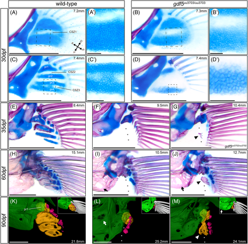
gdf5uu3703/uu3703 mutants display loss of pectoral fin radials. All images depict pectoral fins with postcleithra removed in a dorsomedial view. Bone- and cartilage-stained pectoral fins of wild-type and gdf5uu3703/uu3703 zebrafish at 30 dpf (A-D), 35 dpf (E-G), and 60 dpf (H-J). Dashed boxes (A-D) mark magnified regions (A′-D′). (F,G) pectoral fins of two 35 dpf gdf5uu3703/uu3703 and gdf5uu3702/uu3702 zebrafish, respectively, and (I,J) pectoral fins of two 60 dpf gdf5uu3703/uu3703 zebrafish, displaying variable presence of bone at the position of proximal radial 2 or 3. (K) pectoral fin of a 90 dpf nonsibling wild-type zebrafish following μCT scanning and 3D segmentation. (L,M) pectoral fin of 90 dpf gdf5uu3703/uu3703 zebrafish following μCT scanning and 3D segmentation. The measurements in mm refer to SL. Asterisks highlight the absence of proximal radials 2-4. Arrowheads indicate the variably present bone in place of proximal radial 2 in gdf5uu3703/uu3703 fish. Arrows indicate increased porosity in the shoulder girdle of gdf5uu3703/uu3703 fish. In μCT images, the shoulder girdle (green) is rendered partially transparent to provide unobstructed views of the proximal radials (orange). Fin rays (white) were removed for clarity and shown instead in the insets. Distal radials are colored in pink. CSZ, cartilage segmentation zone; pr, proximal radial. Scale bars: 500 μm (A-M), 50 μm (A′-D′)
|
Expression Data
Expression Detail
Antibody Labeling
Phenotype Data
| Fish: | |
|---|---|
| Observed In: | |
| Stage Range: | Days 30-44 to Adult |
Phenotype Detail
Acknowledgments
This image is the copyrighted work of the attributed author or publisher, and
ZFIN has permission only to display this image to its users.
Additional permissions should be obtained from the applicable author or publisher of the image.
Full text @ Dev. Dyn.