
Fig. 3
- ID
- ZDB-FIG-220804-3
- Publication
- Zhang et al., 2022 - Inner nuclear membrane protein TMEM201 maintains endothelial cell migration and angiogenesis by interacting with the LINC complex
- Other Figures
- All Figure Page
- Back to All Figure Page
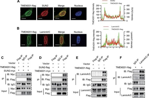
TMEM201 interacts with LINC complex components SUN2 and LaminA/C in EC. (A and B) TMEM201-flag was detected by immunofluorescence in HUVECs with anti-Flag antibody and costained for SUN2 (A) or LaminA/C (B). The dotted yellow line was examined for the colocalization analysis, shown on the right. The X-axis shows the relative position of the dotted yellow line. The Y-axis shows the intensity signals in the green and red channels. Scale bar, 10 μm. (C) CoIP assay and western blotting analysis show that TMEM201-flag coimmunoprecipitated with SUN2-myc. EA.hy926 cells were transfected with vector or TMEM201-flag together with SUN2-myc. Cell lysates were immunoprecipitated with anti-Flag or control IgG and immunoblotted with antibodies against Flag and Myc. (D) CoIP assay and western blotting analysis show that SUN2-flag coimmunoprecipitated with TMEM201-myc. EA.hy926 cells were transfected with vector or SUN2-flag together with TMEM201-myc. Cell lysates were immunoprecipitated with anti-Flag or control IgG and immunoblotted with antibodies against Flag and Myc. (E) CoIP assay and western blotting analysis show that TMEM201-flag coimmunoprecipitated with endogenous LaminA/C. EA.hy926 cells were transfected with vector or TMEM201-flag. Cell lysates were immunoprecipitated with anti-Flag or control IgG and immunoblotted with antibodies against Flag and LaminA/C. (F) CoIP assay and western blotting analysis show that endogenous LaminA/C coimmunoprecipitated with TMEM201-flag. EA.hy926 cells were transfected with TMEM201-flag. Cell lysates were immunoprecipitated with anti-LaminA/C or control IgG and immunoblotted with antibodies against Flag and LaminA/C. |