FIGURE

FIGURE 1

ID
ZDB-FIG-220406-32
Publication
Lv et al., 2022 - Tcap Deficiency in Zebrafish Leads to ROS Production and Mitophagy, and Idebenone Improves its Phenotypes
Other Figures
All Figure Page
Back to All Figure Page
FIGURE 1

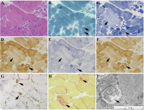
The muscle biopsy specimen from LGMD2G patient. (A) HE staining showed obvious variation in fiber size. (B) MGT staining showed scattered rimmed vacuoles. No ragged red fiber was found in MGT staining. (C–E) The presence of high enzyme activities for NADH (C), COX (D), and SDH (E) staining at the periphery of sarcoplasm and the decreased enzyme activities in the center of sarcoplasm. (F) No blue fibers in SDH-COX double staining. (G) P62 staining showed scattered P62-positive muscle fibers. (H) Increased acid phosphatase enzymatic activity (black arrows). (I) Ultrastructural examination and muscle pathology of patients with LGMD2G.

Expression Data

Expression Detail
Antibody Labeling
Phenotype Data

Phenotype Detail
Acknowledgments
This image is the copyrighted work of the attributed author or publisher, and ZFIN has permission only to display this image to its users. Additional permissions should be obtained from the applicable author or publisher of the image. Full text @ Front Cell Dev Biol