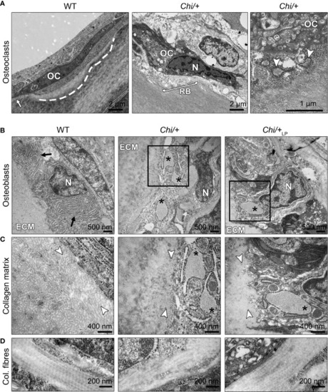
Ultrastructure of bone cells and bone matrix in Chi/+ and Chi/+LP animals. (A) Transmission electron microscopy (TEM) of osteoclasts (OC) located at the arch surface of three months old WT and Chi/+ (representative for both Chi/+ and Chi/+LP) vertebrae. The panel WT shows a typical flat-shaped teleost osteoclast (OC) characterised by its electrodense cytoplasm. The cell resides in a shallow resorption lacuna (dashed line), yet is attached to the bone surface (white arrow). The panel Chi/+ shows an osteoclast that resorbs the bone matrix and exhibits the typical ‘ruffled border’ (RB), an electron-dense cytoplasm with abundant cytoplasmic vacuoles and resorption vesicles (right panel, white arrowheads) in proximity of the ruffled border. N, nucleus. (B) TEM of WT, Chi/+ and Chi/+LP shows mutant osteoblasts with enlarged endoplasmic reticulum (ER) cisternae (asterisks). The osteoblasts are located in the growth zone of the vertebral body endplate (see Figure 3A for location). Higher magnification images in (C) show that Chi/+ osteoblast ER cisternae are filled with protein, likely mutated collagen type I WT osteoblasts have numerous, yet not enlarged ER cisternae (arrows). ECM: extracellular matrix of the bone surface; N: nucleus. (C) The newly secreted collagen fibrils (white arrowheads) in the proximity of the osteoblasts in WT are visibly separated prior to maturation and assemblage into collagen fibres. In contrast, the Chi/+ animal has densely packed collagen fibrils. No space can be recognised between the fibrils. In the Chi/+LP individual collagen fibres are densely packed and space between the fibrils is distinguishable in proximity to the osteoblasts. (D) Longitudinal sections of collagen fibres show a regular D-periodicity pattern in a WT animal, absence D-period pattern in a Chi/+ mutant, and a less regular D-periodicity pattern a Chi/+LP specimen.
|
