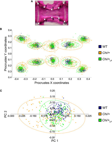
Vertebral body shape variation in Chi/+ and Chi/+LP mutants. (A) Alizarin red S stained vertebral body of a WT animal with 2D landmark positions used for quantifying the shape variation by means of geometric morphometrics, represented in (B). (B) The scatterplot of WT, Chi/+ and Chi/+LP 2D landmarks shows high variation in the superimposition of X,Y Procrustes coordinates of Chi/+ compared to WT animals. Chi/+LP animals display reduced landmark variation compared to Chi/+ and a distribution more similar to WT indicating a partial rescue of shape variation at three months of age. The first 10 caudal vertebral centra in WT (n=15), Chi/+ (n=13) and Chi/+LP (n=12) were analysed. The 95% confidence ellipses are shown. (C) Principal component analysis of vertebral centra shapes. Each symbol in the plot represents a vertebral body. PC indicates Principal Component and the values in the axis labels indicate the percentage of variation accounted for by each axis. Chi/+ animals show high variance compared to WT animals (Chi/+ versus WT, PC1 0.3321, PC2 0.1795; Chi-square test: p < 0.001). Variance is rescued in Chi/+LP animals (Chi/+LP versus WT, PC1 0.2484, PC2 0.2020; Chi-square test: non-significant). The 95% confidence ellipses are shown.
|
