Figure 1—figure supplement 1.

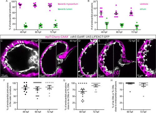
Endocardial protrusions in the ventricle are mostly located in the outer curvature and are close to delaminating or trabecular cardiomyocytes (CMs).

(A) Quantification of the direction of endocardial protrusions; most endocardial protrusions extend toward the myocardium. (B) Quantification of the average number of endocardial protrusions in the ventricle and atrium. (C–D) Confocal projection images of the heart of Tg(myl7:mCherry-CAAX); Tg(cdh5:Gal4ff); Tg(UAS:LIFEACT-GFP) zebrafish at 60 (C) and 72 (D) hours post-fertilization (hpf). Arrows point to endocardial protrusions; arrowheads point to delaminating CMs and trabecular CMs close to endocardial protrusions. (E) Quantification of the percentage of endocardial protrusions in the outer curvature of the ventricle. (F) Quantification of the percentage of endocardial protrusions in close proximity to delaminating CMs or trabecular CMs. (G) Quantification of the percentage of delaminating CMs or trabecular CMs in close proximity to endocardial protrusions. de CM, delaminating CM; TL CM, trabecular layer CM.

Expression Data

Expression Detail
Antibody Labeling
Phenotype Data

Phenotype Detail
Acknowledgments
This image is the copyrighted work of the attributed author or publisher, and ZFIN has permission only to display this image to its users. Additional permissions should be obtained from the applicable author or publisher of the image. Full text @ Elife