
Fig. 1.
- ID
- ZDB-FIG-220304-10
- Publication
- Torregrosa-Carrión et al., 2021 - Adhesion G protein-coupled receptor Gpr126/Adgrg6 is essential for placental development
- Other Figures
- All Figure Page
- Back to All Figure Page
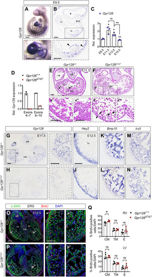
Defective chamber development in Gpr126Δ7/Δ7 mutant embryos is not associated with altered patterning or proliferation.
(A) E9.5 WT embryo. Whole-mount in situ hybridization (WISH) showing Gpr126 mRNA in somites (arrowheads) and heart (bottom). (B) Transverse heart sections. Bottom: Arrowheads indicate endocardial Gpr126 expression. (C) Quantitative reverse transcription polymerase chain reaction (qRT-PCR): Gpr126 transcription (relative to Gapdh) in embryonic WT hearts. Data are means ± SD (n = 3 pools of three hearts per pool per stage). Relative values normalized to E9.5. Statistics: Unpaired Student’s t test (ns, not significant; *P < 0.05; **P < 0.01). (D) qRT-PCR. Gpr126 gene expression (relative to Gapdh) in Gpr126 +/+ and Gpr126Δ7/Δ7 E12.5 hearts, using primers spanning exons 4 to 7 and exons 9 and 10. Data are means ± SD (n = 2 hearts). (E to F″) Hematoxylin and eosin (H&E)–stained E11.5 Gpr126+/+ (E to E″) and Gpr126Δ7/Δ7 heart sections (F to F″). Bottom panels: High magnifications of chamber (′) and septum (″). Note compact myocardium (arrowheads) and trabeculae (arrows) thinning and poorly cellularized ventricular septum (asterisk) in the mutant (F′ and F″). (G and H) Gpr126 ISH, E11.5 Gpr126+/+ (G) and Gpr126Δ7/Δ7 (H) heart sections. Right: Higher magnifications. (I to N) ISH of compact myocardium [Hey2 (I and J)], trabecular myocardium [Bmp10 (K and L)], and chamber endocardium [Irx5 (M and N)] markers in E12.5 Gpr126+/+ and Gpr126Δ7/Δ7 hearts. (O to P″) BrdU immunostaining (red) of E12.5 Gpr126+/+ (O to O″) and Gpr126Δ7/Δ7 (P to P″) hearts. α-Smooth muscle actin (α-SMA) (green) counterstains the myocardium, ERG (white) counterstains the endocardium, and 4′,6-diamidino-2-phenylindole (DAPI) (blue) counterstains the nuclei. The panels (′ and ″) show high magnifications of boxed areas in (O) and (P). (Q) BrdU-positive nuclei quantification as a percentage of total nuclei (DAPI+) in the compact (CM), trabecular myocardium (TM), and endocardium (E) of E12.5 Gpr126+/+ and Gpr126Δ7/Δ7 hearts. Data are means ± SD. Statistics: Unpaired Student’s t test. Scale bars, 100 μm (low magnification) and 50 μm (high magnification). a, atrium; avc, atrioventricular canal; ivs, interventricular septum; la, left atrium; lv, left ventricle; oft, outflow tract; ra, right atrium; rv, right ventricle. |