
Fig. 5
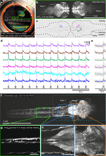
Mesoscale imaging of zebrafish larvae.
a Photograph of two 9 dpf zebrafish larvae mounted head-to-head in a microscope chamber with mm-scale ruler in background. b The same 2 fish (HuC:GCaMP6f) as in (a) imaged under 2-photon with nTC2 3.5 mm FOV configuration, at 512 × 128 px (3.91 Hz). c, d Activity-correlation (cf. Fig. 3c) of the scan in (b) during presentation of full-field flashes of ultraviolet (UV)-light, with hand-selected exemplary ROIs, extracted time-traces (d) and light-stimulus-aligned averages (e). f–i The same fish as shown on the left in (b, fish 1), now shown at full 3.5 mm field of view (f, 512 × 128 px, 3.91 Hz) and increased spatial resolution scans of regions as indicated to reveal cellular detail (g–i, 1024 × 1024 px, 0.49 Hz). RGC, retinal ganglion cell. |