
Fig. 7
- ID
- ZDB-FIG-220203-95
- Publication
- Missinato et al., 2021 - Zebrafish heart regenerates after chemoptogenetic cardiomyocyte depletion
- Other Figures
- All Figure Page
- Back to All Figure Page
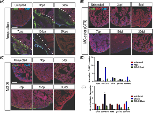
Myofibroblasts are mildly activated upon cardiomyocyte chemoptogenetic ablation. A, Activated fibroblasts (αSMA) and MHC (cardiac muscle) immunostaining before and after ventricular amputation at several time points. In uninjured ventricles αSMA staining is not detectable. After amputation, αSMA staining is visible inside the injury but by 30 dpa activated fibroblasts were not detected. B, Tg(myl7:fapdl5-cerulean) hearts injected with control MG-ester and treated with near IR-light, stained for αSMA and MHC. αSMA was not detected at any time point. C, In chemoptogenetic ablated hearts, myofibroblasts are weakly activated starting at 5 dpi, until 15 dpi. At least n = 3 for each time point. Q-PCR of cytochrome b, (cybb), collagen (col12a1a and col1a1b), fibronectin (fn1b), and periostin a (postna) in amputated and ablated hearts at 7 days and 5 days (D) and at 20 days (E). Scale bar = 100 μm |