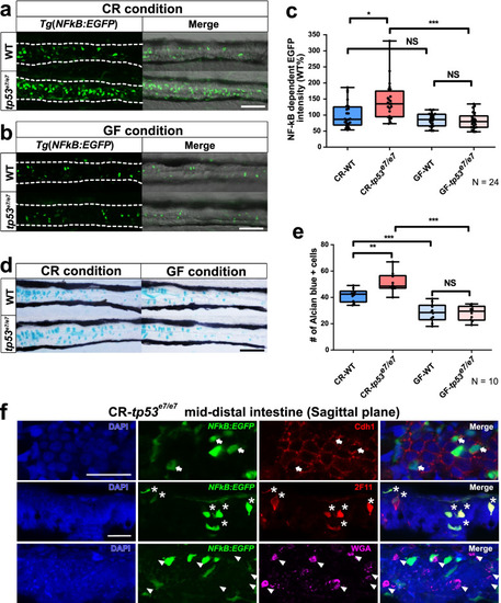
tp53 mutants exhibit elevated NF-κB signaling in the mid-distal intestines due to intestinal bacteria. a, b NF-κB signaling in the mid-distal intestines of WT or tp53 mutants was monitored using Tg(NFκB:EGFP) transgenic zebrafish larvae at 7 dpf. CR (a), conventionally raised. GF (b), germfree. Scale bar = 50 μm. White dashed lines denote boundaries of GITs based on DIC bright field images. c A boxed plot showing comparison of fluorescence intensity of the mid-distal intestines of WT and tp53 mutants under CR- or GF-condition. N = 24 each. d Alcian blue staining visualizing goblet cells in the mid-distal intestines of WT or tp53 mutants under CR- or GF- conditions. N = 10 each. Scale bar = 50 μm. e A boxed plot showing comparison of the number of Alcian blue-positive goblet cells in the mid-distal intestines of WT and tp53 mutants under CR- or GF- conditions. f To reveal cell types of NF-κB positive cells, EGFP-positive cells in the sagittal sections through the mid-distal intestine of tp53 mutant Tg(NFκB:EGFP) at 7dpf under CR condition stained for markers of pan-secretory cells (2F11), enterocytes (Cdh1), goblet cells (WGA) using immunohistochemistry. The NF-κB dependent EGFP-positive intestinal cells were clearly positive with Cdh1 (white arrows) and 2F11 (white asterisks), but negative with WGA (white arrowheads)-positive cells. Scale bar = 20 μm. The boxed plots in (c) and (e) were statistically estimated by non-parametric Friedman test followed by Dunn’s multiple comparisons test. Data are represented as mean ± SEM. *< p = 0.05; **< p = 0.01; ***< p = 0.005
|
