
FIGURE
Figure 5
- ID
- ZDB-FIG-210801-73
- Publication
- Chuang et al., 2021 - Oxytocin Signaling Acts as a Marker for Environmental Stressors in Zebrafish
- Other Figures
- All Figure Page
- Back to All Figure Page
Figure 5
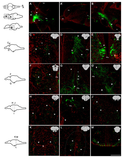
Distribution and projection of oxytocin neurons in the adult zebrafish brain (n = 5). Whole-brain sections were stained with anti-GFP (green; oxytocin) and Nissl (red). (A,B) Sagittal sections showed that the fibers of oxytocin neurons were widely present in the brain. Somas were found in (D,E) PPa, (F,G) PPp, and (I) TPp. From the rostral end to the caudal end, the regions containing the fibers were found in (C) Vv, (D,E) PPa, (F) PPp, (G) A, VM, (H) PGm, (I) TPp, (J) Cans, (K) IPN, (L) raphe, and (M) TTBc. Scale bar = 100 µm. |
Expression Data
Expression Detail
Antibody Labeling
Phenotype Data
Phenotype Detail
Acknowledgments
This image is the copyrighted work of the attributed author or publisher, and
ZFIN has permission only to display this image to its users.
Additional permissions should be obtained from the applicable author or publisher of the image.
Full text @ Int. J. Mol. Sci.