|
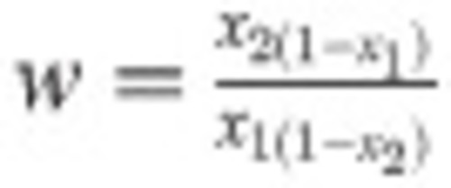
The role of PBP4 in <italic>S</italic>. <italic>aureus</italic> virulence.Mice (n = 10) were injected with approximately 5x10⁶ CFU S. aureus NewHG kanR (WT, SJF 3680) or NewHG pbp4::ery (SJF 5103). (A) Weight loss 72 hpi and CFUs recovered from (B) livers, (C) kidneys were determined. Groups were compared using a Mann-Whitney U test (NewHG kanR–black circles, NewHG pbp4::ery blue squares) (** p = 0.0051, * p = 0.0410). (D) Mice (n = 20) were injected with a 1:1 ratio (totalling 7 x 10⁶ CFU) of NewHG kanR (SJF 3680, red) and NewHG pbp4::ery (SJF 5103, blue). 72 hpi mice were culled and CFU ratios within organs were determined. The number in each pie chart represents the log total number of bacteria recovered (i.e. 106 CFU = 6). Mice 9 and 12 were culled at 54 hpi due to severity limits but are included in the analysis. Mouse 18 was found dead at 72 hpi and is excluded from analysis. The relative fitness of NewHG pbp4::ery (SJF 5103) against NewHG kanR (SJF 3680) was calculated using the formula ![]() w=x2(1−x1)x1(1−x2) (where w = relative fitness, X1= starting mutant proportion and X2= ending mutant proportion). This was calculated for the (E) liver (* p = 0.0105), (F) left kidney (p = 0.3258), (G) right kidney (* p = 0.0327), (H) spleen (* p = 0.0476), (I) lungs (p = 0.3396) and (J) heart (p = 0.43275). Line on graph depicts the median. Statistical significance was determined using a one sample Wilcoxon signed rank test, comparing the results to a theoretical median of 1, which would indicate an equal fitness between the strains. w=x2(1−x1)x1(1−x2) (where w = relative fitness, X1= starting mutant proportion and X2= ending mutant proportion). This was calculated for the (E) liver (* p = 0.0105), (F) left kidney (p = 0.3258), (G) right kidney (* p = 0.0327), (H) spleen (* p = 0.0476), (I) lungs (p = 0.3396) and (J) heart (p = 0.43275). Line on graph depicts the median. Statistical significance was determined using a one sample Wilcoxon signed rank test, comparing the results to a theoretical median of 1, which would indicate an equal fitness between the strains.
|