
Fig. 5
- ID
- ZDB-FIG-210129-26
- Publication
- Ghilardi et al., 2020 - Expression pattern of the small muscle protein, X-linked (smpx) gene during zebrafish embryonic and larval developmental stages
- Other Figures
- All Figure Page
- Back to All Figure Page
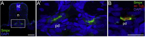
Smpx localizes to the ciliated cells of the zebrafish pronephros. (A) Cross-section of the trunk of a 48 hpf embryo; the pronephric ducts are labeled in green with the antibody against Smpx. (A′) 5X magnifications of the area enclosed in the rectangle in A; Smpx labels the apical membrane of the ciliated cells facing the lumen of the duct, as also confirmed (B) by the co-labeling with antibodies against Smpx and acetylated tubulin, where the lumen is populated exclusively by the acetylated tubulin-based kinocilia (red) sprouting from the apical membrane and devoid of Smpx (green). Images are all confocal Z-stacks taken from paraffin sections. pd, pronephric duct; n, notochord; nt, neural tube. Scale bars = 50 μm in A; 20 μm in C. |
| Gene: | |
|---|---|
| Antibody: | |
| Fish: | |
| Anatomical Terms: | |
| Stage: | Long-pec |
Reprinted from Gene expression patterns : GEP, 36, Ghilardi, A., Diana, A., Prosperi, L., Del Giacco, L., Expression pattern of the small muscle protein, X-linked (smpx) gene during zebrafish embryonic and larval developmental stages, 119110, Copyright (2020) with permission from Elsevier. Full text @ Gene Expr. Patterns