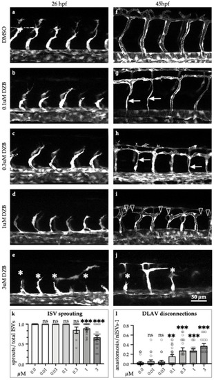
Derazantinib inhibits vascular development in vivo in a dose-dependent manner. Confocal images of GFP+ blood vessels in the trunk of Tg(fli1:EGFP)y1 zebrafish embryos at 26 hpf (a–e) or 45 hpf (f–j) after exposure to DMSO as vehicle control (a and f) or increasing concentrations of DZB in the swimming water (b–e and g–j). Blood vessel development was disrupted using concentrations between 0.1 and 3 µM DZB. (k) Quantitative analysis of ISV sprouts that had reached the top roof and started to form the DLAV were normalised to the total number of ISVs per embryo (n ≥ 15 embryos per treatment were analysed from three independent experiments), embryos were treated using increasing concentrations of DZB. (l) Quantitative analysis of ISV sprouts that are disconnected at the DLAV were normalised to the total number of ISVs-1 (total number of connections) per embryo (n ≥ 15 embryos per treatment were analysed from three independent experiments) and embryos were treated using increasing concentrations of DZB. Data in k,l represent mean ± S.E.M. (error bars), ns: not significant, ** p < 0.01, *** p < 0.001. Arrows indicate thinner blood vessels, arrowheads show disconnected blood vessels and asterisks mark sprouting defects. Scale bar, 50 µm. ISV, intersegmental vessel; DLAV, dorsal longitudinal anastomotic vessel; DA, dorsal aorta; PCV, posterior cardinal vein. See also Figure S1.
|
