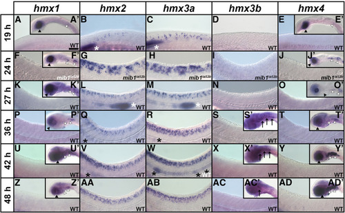
Expression of hmx genes in WT zebrafish embryos. Lateral views of hmx expression in spinal cord (A–AD), hindbrain (S’, X’, AC’), eye and ear (A’, E’, F’, J’, K’, O’, P’, T’, U’, Y’, Z’, AD’), and lateral line primordium and neuromasts (B, C, L, M, Q, R, V, W) at 19 hpf (A–E, A’, E’), 24 hpf (F–J, F’, J’), 27 hpf (K–O, K’, O’), 36 hpf (P–T, P’, S’, T’), 42 hpf (U–Y, U’, X’, Y’), and 48 hpf (Z–AD, Z’, AC’, AD’). Rostral, left; Dorsal, up. (B, C, G, H, L, M, Q, R, V, W, AA, AB) hmx2 and hmx3a are expressed in spinal cord, lateral line primordium (white asterisks), neuromasts (black asterisks), and anterior ear (data not shown) at all stages examined, although hmx2 spinal cord expression initially appears weaker than hmx3a and does not extend as far caudally. While there is expression of both hmx2 and hmx3a in the lateral line primordium at 24 hpf (data not shown), the lateral line primordium has not yet migrated into the field of view shown in G and H. For consistency, the specific region of spinal cord shown (adjacent to somites 6–10) is identical in panels F–AD. At 19 hpf, expression is found only in the very anterior spinal cord and so a more rostral region of spinal cord is shown in A–E. (A, A’, E, E’, F, F’, J, J’, K, K’, O, O’, P, P’, T, T’, U, U’, Y, Y’, Z, Z’, AD, AD’) hmx1 and hmx4 are not expressed in WT spinal cord at any of these stages but are expressed in the eye (black arrowheads), and posterior-ventral ear and adjacent ganglion of the anterior lateral line (white arrowheads). (D, I, N, S, S’, X, X’, AC, AC’) hmx3b is not expressed in WT spinal cord at any of these stages. The only expression we observed was in the hindbrain between 36 and 48 hpf (black arrows). (G and H) The expression pattern of hmx2 and hmx3a is expanded in the spinal cord of mib1ta52b mutants but is unaltered in the ear and lateral line primordium (data not shown). (F, F’, J, J’) Neither hmx1 (F) nor hmx4 (J) are expressed in the spinal cord of mib1ta52b mutants, although the expression of both genes persists in the eye (black arrowheads), posterior-ventral ear and adjacent ganglion of the anterior lateral line (white arrowheads) (F’ and J’). (I) hmx3b is not expressed in mib1ta52b mutants, either in the spinal cord or in any other tissue. (F, I, J, K, N, O, P, S, S’, T, U’, X, X’, Y, Z, AC, AC’, AD) The background (diffuse, nonspecific staining) in these pictures is higher because we exposed the embryos to prolonged staining to ensure that there was no weak spinal cord expression. Especially in the brain, this can lead to background staining as the large ventricles of the hindbrain trap anti-sense riboprobes. Bar, 50 µm (A–AD), 120 µm (A’, E’, F’, J’, K’, O’, P’, S’, T’, U’, X’, Y’, Z’, AC’, AD’).
|
