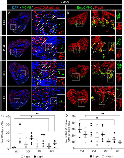
A decreased activation of cardiomyocyte proliferation and dedifferentiation after multiple cryoinjuries. (A) Cross sections of transgenic zebrafish hearts at 7 dpci following 1, 2, 3 or 6 CIs expressing nuclear DsRed in cardiomyocytes. Proliferating cells are detected by immunostaining against Minichromosome Maintenance Complex Component 5 (MCM5; green). Proliferating cardiomyocytes are observed (white arrows) by colocalization between cmlc2:DsRed2-nuc and MCM5 in the vicinity of the wound (encircled with a dash line). Scale bars = 50 µm. (B) Cross sections of adult zebrafish hearts at 7 dpci following 1, 2, 3 or 6 CIs, immunostained for embryonic cardiac myosin heavy chain (EmbCMHC; N2.261; green). The cardiac muscle is detected by F-actin staining (Phalloidin, red). EmbCMHC-positive cardiomyocytes are detected in the peri-injury zone within an area of 100 µm from the injury border (dashed line). (C) Analysis of the percentage of MCM5-positive nuclei of cardiomyocytes (CMs) at 4 and 7 dpci, following multiple cryoinjuries. N ≥ 4. *P < 0.05; **P < 0.01. Scatter plot of the data with a large bar indicating the mean and smaller bars representing the SEM. (D) Analysis of the percentage of embCMHC-positive cardiomyocytes (CMs) at 7 and 14 dpci, within the peri-injury zone. N ≥ 4. **P < 0.01. Scatter plot of the data with a large bar indicating the mean and smaller bars representing the SEM.
|
