FIGURE

Figure 1

ID
ZDB-FIG-200718-17
Publication
Bise et al., 2020 - Multiple cryoinjuries modulate the efficiency of zebrafish heart regeneration
Other Figures
All Figure Page
Back to All Figure Page
Figure 1

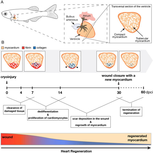
Schematic summary of the regenerative processes after cryoinjury in the zebrafish heart. (A) Illustration of the anatomy and histology of an intact zebrafish heart. The ventricle comprises a trabecular myocardium (beige) that is surrounded by a thin layer of a compact myocardium (orange). (B) Illustration of the main cellular processes after cryoinjury during the regeneration time. The prominent events are written in the boxes and linked to the specific periods after cryoinjury. The schematic heart sections were based on AFOG histological staining that visualizes the myocardium in beige/orange, collagen in blue and fibrin-like material in red. The injury zone switches from red staining at 4 and 7 dpci, to blue collagen staining after 7 dpci. At the bottom, the rectangular graph depicts a progressive replacement of the wound with a new myocardium. During this time, the wounded tissue undergoes remodeling, starting from the inflammatory state (red gradient) followed by collagenous scar deposition (inverse blue gradient).

Expression Data

Expression Detail
Antibody Labeling
Phenotype Data

Phenotype Detail
Acknowledgments
This image is the copyrighted work of the attributed author or publisher, and ZFIN has permission only to display this image to its users. Additional permissions should be obtained from the applicable author or publisher of the image. Full text @ Sci. Rep.