FIGURE

Figure 6

ID
ZDB-FIG-200713-16
Publication
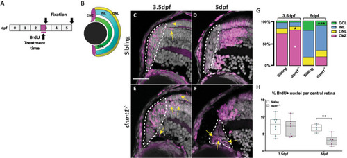
Angileri et al., 2020 - dnmt1 function is required to maintain retinal stem cells within the ciliary marginal zone of the zebrafish eye
Other Figures
All Figure Page
Back to All Figure Page
Figure 6

Neurons produced by dnmt1−/− RSCs fail to integrate into the neural retina. A Experimental paradigm depicting BrdU incorporation from 3 to 3.5dpf. Fixations occurred at 3.5 and 5dpf. B Diagram of the four retinal domains (CMZ, GCL, INL, and ONL) whose colors correlate with the data presented in G. CF Transverse sections of BrdU pulses from 3 to 3.5dpf (C,E) and pulse-chase assay from 3 to 5dpf (D, F) (Siblings: A, B; dnmt1−/−C, D). Nuclei labeled with DAPI (gray). Cells in S-phase indicated by BrdU incorporation (magenta). Mitotic cells are labeled by a pH3(ser10) antibody (cyan). G Proportion of BrdU + cells located in each retinal layer at 3.5dpf and 5dpf of dnmt1−/− and control larvae. H Proportion of total BrdU+ cells within the central retina of the pulse-chase experiment. White dotted lines designate the CMZ (CF). Yellow arrows = BrdU+ nuclei outside the CMZ (C, E). Scale bars: 20 μm. *p < 0.05, **p < 0.005, ***p < 0.0005. Dorsal is up in all images.

Expression Data

Expression Detail
Antibody Labeling
Phenotype Data

Phenotype Detail
Acknowledgments
This image is the copyrighted work of the attributed author or publisher, and ZFIN has permission only to display this image to its users. Additional permissions should be obtained from the applicable author or publisher of the image. Full text @ Sci. Rep.