|
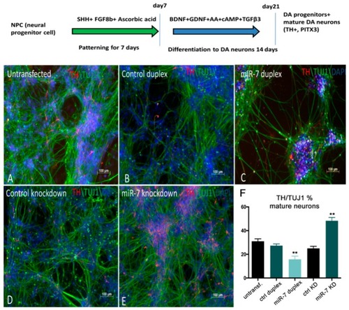
miR-7 negatively regulates the number of TH+ DA neurons derived from H9NPC cells. (A,B,C) Control H9NPC cells (A) showing TH staining at day 10 of DA neurogenesis (schematized in the top of the figure). BDNF: Brain Derived Neurotrophic Factor; GDNF: Glial cell Derived Neurotrophic Factor; cAMP: cyclic Adenosine MonoPhosphate; TGFβ3: Transforming Growth Factor beta 3. Decrease of TH+ cells (red) was observed when cells were transfected with miR-7 duplex (C), compared with the control duplex (B) and untransfected cells (A). (D,E) Increase of TH+ cells was observed upon knockdown of miR-7 (E), when compared with the control antisense. (F) Ratio of TH+/TUJ1 cells was quantified by manual counting of the neurons from images captured from 10 different areas on a random basis. Error bars represent average% ± SEM; the experiment was repeated thrice with n = 3 different biological replicates; (**) p < 0.01.
|