FIGURE

Figure 2

ID
ZDB-FIG-191230-1731
Publication
Simbulan-Rosenthal et al., 2019 - CRISPR-Cas9 Knockdown and Induced Expression of CD133 Reveal Essential Roles in Melanoma Invasion and Metastasis
Other Figures
All Figure Page
Back to All Figure Page
Figure 2

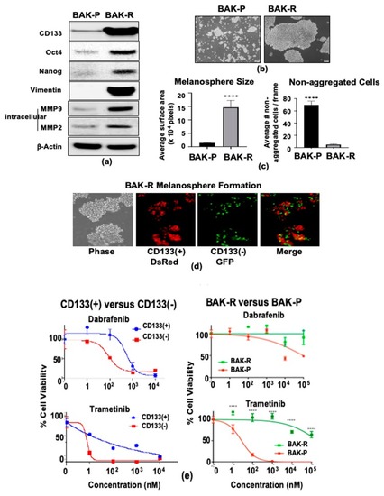
Compared to BAK-P, BAK-R cells strongly express markers of cancer stem cells (Oct4, Nanog), EMT (vimentin), and invasion (MMP2 and MMP9) as shown by immunoblot analysis (a), melanosphere formation (bd) and resistance to kinase inhibitors (e). BAK-P and BAK-R cells were cultured separately in melanosphere conditions, imaged (b) and melanosphere sizes as well as average number of non-aggregated cells were quantified by Image J analysis (c). CD133(+) DsRed and CD133(−) GFP BAK-R cells were recombined in a 1:1 ratio and grown in low attachment plates (d). CD133(+) cells derived by MACS sorting (left column) and BAK-R (right column) both exhibit increased resistance to dabrafenib (top) or trametinib (bottom), compared to CD133(−) cells (left panel) or parental cells (BAK-P; right panel). p-values < 0.0001 are shown as four asterisks (c,e).

Expression Data

Expression Detail
Antibody Labeling
Phenotype Data

Phenotype Detail
Acknowledgments
This image is the copyrighted work of the attributed author or publisher, and ZFIN has permission only to display this image to its users. Additional permissions should be obtained from the applicable author or publisher of the image. Full text @ Cancers