FIGURE

Figure 1

ID
ZDB-FIG-191230-1730
Publication
Simbulan-Rosenthal et al., 2019 - CRISPR-Cas9 Knockdown and Induced Expression of CD133 Reveal Essential Roles in Melanoma Invasion and Metastasis
Other Figures
All Figure Page
Back to All Figure Page
Figure 1

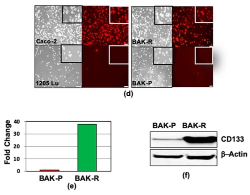
Preferential survival and growth of MACS-sorted CD133(+) cells in mixed population xenografts, and sustained CD133 expression after reprogramming. GFP- CD133(+) and DsRed-CD133(−) cells were isolated by MACS (a), injected into the flanks of nude mice, and tumors were visualized and measured by Maestro in vivo imaging after indicated periods of time. p-values of <0.05 were considered statistically significant and represented with a single asterisk, while p < 0.01 are labeled with two asterisks. (b). (c) CD133 expression of reprogrammed BAK-R MIC and BAK-P populations assessed by immunofluorescent staining with anti-CD133-PE for 3 weeks show that BAK-R, but not BAK-P melanoma cells, exhibit sustained CD133 expression. Representative images after immunofluorescent staining of BAK-P and BAK-R cells with anti-CD133-PE (d, right panels) as well as qRT-PCR (e) and immunoblot (f) analyses showing elevated CD133 expression in BAK-R compared to BAK-P cells. CD133(+) Caco-2 colon cancer cells and CD133(−) 1205Lu melanoma cells served as positive and negative controls, respectively (d; left panels).

Expression Data

Expression Detail
Antibody Labeling
Phenotype Data

Phenotype Detail
Acknowledgments
This image is the copyrighted work of the attributed author or publisher, and ZFIN has permission only to display this image to its users. Additional permissions should be obtained from the applicable author or publisher of the image. Full text @ Cancers