
Fig. 5
- ID
- ZDB-FIG-180911-45
- Publication
- Oosterhof et al., 2018 - Colony-Stimulating Factor 1 Receptor (CSF1R) Regulates Microglia Density and Distribution, but Not Microglia Differentiation In Vivo
- Other Figures
- All Figure Page
- Back to All Figure Page
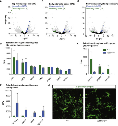
Differential Gene Expression of csf1r-Deficient Microglia Shows Normal Microglia Differentiation (A) Volcano plot showing expressional changes of the 300 most highly expressed microglia-specific genes in csf1ra−/−;b+/− mutant microglia (Oosterhof et al., 2017). (B) Volcano plot showing the expressional changes in csf1ra−/−;b+/− mutant microglia of normally downregulated genes during differentiation (Matcovitch-Natan et al., 2016) and of genes normally expressed in other macrophages in the CNS (Bennett et al., 2016). (C) Volcano plot showing the expression changes of non-microglia myeloid genes. (D) Expression values of zebrafish microglia-specific genes. (E) Expression values of downregulated microglia-specific genes. (F) Expression values of upregulated microglia-specific genes. (G) Representative images of microglia (5-month-old fish) in the ventral part of the optic tectum labeled with an antibody against L-plastin. Scale bar, 20 μm. |
| Antibody: | |
|---|---|
| Fish: | |
| Anatomical Term: | |
| Stage: | Adult |