
Fig. 3
- ID
- ZDB-FIG-180620-48
- Publication
- Cai et al., 2018 - Knockout of zebrafish interleukin 7 receptor (IL7R) by the CRISPR/Cas9 system delays retinal neurodevelopment
- Other Figures
- All Figure Page
- Back to All Figure Page
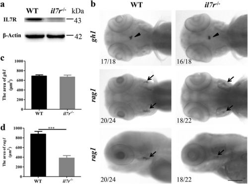
The validation of the il7r−/− mutant by western blotting and whole-mount in situ hybridisation. a The expression of IL7R protein in WT and il7r−/− larvae at 4 dpf. Note that no IL7R protein is detected in il7r−/− larvae. b The images of whole-mount in situ hybridisation with gh1 (arrowheads) and rag1 (arrows) mRNA probes in WT and il7r−/− larvae at 4 dpf. c-d The statistical analysis of gh1-positive area (c) or rag1-positive area (d) between WT and il7r−/− larvae. Note that rag1 expression is significantly decreased in il7r−/− larvae. Results are represented as means ± SEM, ***P < 0.001. The upper four panels in b: dorsal view. The lower two panels in b: dorsal is up, and rostral is left. Scale bar in b: 100 μm |
| Genes: | |
|---|---|
| Fish: | |
| Anatomical Terms: | |
| Stage: | Day 4 |
| Fish: | |
|---|---|
| Observed In: | |
| Stage Range: | Protruding-mouth to Day 4 |