
Fig. 1
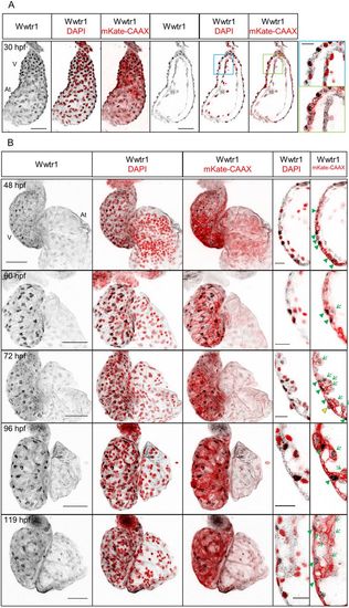
Expression and localization of Wwtr1 in the developing zebrafish heart. (A) Maximum intensity projections and confocal sagittal sections of a whole-mount zebrafish heart at 30 hpf. Magnified images (blue and green squares) show that the nuclei of cardiomyocytes, but not of endocardial cells, are positive for Wwtr1 expression. (B) Maximum intensity projections and confocal sagittal sections of whole-mount zebrafish hearts from 48 to 119 hpf. Wwtr1 is predominantly localized in the nucleus in some ventricular cardiomyocytes (green arrowheads) and moderately expressed in some epicardial cells (yellow arrowhead), but absent from endocardial cells. During trabeculation, some cardiomyocytes delaminate from the compact layer and exhibit weaker nuclear staining for Wwtr1 compared with adjacent compact layer cardiomyocytes (green arrows and Fig. S1B). Nuclei are counterstained with DAPI and cardiomyocyte membranes are marked with myl7:mKate-CAAX expression. Scale bars: 50 μm; 15 μm (insets). V, ventricle; At, atrium. 30-72 hpf, n>3. 96 and 119 hpf, n=2. |
| Genes: | |
|---|---|
| Antibody: | |
| Fish: | |
| Anatomical Terms: | |
| Stage Range: | Prim-15 to Day 5 |